Instances of traumatic force applied to the chest are seen in 10% of the cases of pediatric trauma, usually in the context of motor vehicle accidentsMotor Vehicle AccidentsSpinal Cord Injuries and falls. Chest trauma rarely occurs in isolation and is often associated with polytraumaPolytraumaMultitrauma occurs when 2 or more traumatic injuries occur in at least 2 areas of the body. A systematic management approach is necessary for individuals who have undergone trauma to maximize outcomes and reduce the risk of undiscovered injuries.Multitrauma. The 2 major mechanisms involve blunt and penetrating forces. PneumothoraxPneumothoraxA pneumothorax is a life-threatening condition in which air collects in the pleural space, causing partial or full collapse of the lung. A pneumothorax can be traumatic or spontaneous. Patients present with a sudden onset of sharp chest pain, dyspnea, and diminished breath sounds on exam.Pneumothorax, hemothoraxHemothoraxA hemothorax is a collection of blood in the pleural cavity. Hemothorax most commonly occurs due to damage to the intercostal arteries or from a lung laceration following chest trauma. Hemothorax can also occur as a complication of disease, or hemothorax may be spontaneous or iatrogenic. Hemothorax, flail chestFlail chestFlail chest is a life-threatening traumatic injury that occurs when 3 or more contiguous ribs are fractured in 2 or more different locations. Patients present with chest pain, tachypnea, hypoxia, and paradoxical chest wall movement. Flail Chest, and lung contusions are the most common injuries. Treatment of affected children is very similar to that of adults, but unique pediatric pathoanatomy dictates important differences in approach and management.
Blunt trauma: an injury caused by the application of a mechanical force by a blunt force or object
Penetrating trauma: an injury caused by a cutting or piercing instrument that interrupts the continuity of tissues
Epidemiology
Infrequent: 10% of pediatric trauma
Rarely occurs in isolation, up to 85% of cases are associated with polytraumaPolytraumaMultitrauma occurs when 2 or more traumatic injuries occur in at least 2 areas of the body. A systematic management approach is necessary for individuals who have undergone trauma to maximize outcomes and reduce the risk of undiscovered injuries.Multitrauma
7%–8% of cases are due to abuse and usually occur in individuals < 3years of age.
Blunt trauma:
85% of cases due to blunt trauma
> 50% of cases of blunt thoracic trauma are associated with head, abdomen, and limb injuries.
Penetrating trauma: more often due to gunshot woundsGunshot woundsDisruption of structural continuity of the body as a result of the discharge of firearms.Penetrating Chest Injury
Flexible ribsRibsA set of twelve curved bones which connect to the vertebral column posteriorly, and terminate anteriorly as costal cartilage. Together, they form a protective cage around the internal thoracic organs.Chest Wall: Anatomy:
Transmission of trauma energy to the lung parenchyma → lung contusions
Unlikely to fractureFractureA fracture is a disruption of the cortex of any bone and periosteum and is commonly due to mechanical stress after an injury or accident. Open fractures due to trauma can be a medical emergency. Fractures are frequently associated with automobile accidents, workplace injuries, and trauma.Overview of Bone Fractures: Fractures indicate high-energy mechanisms and internal injury.
The mediastinumMediastinumThe mediastinum is the thoracic area between the 2 pleural cavities. The mediastinum contains vital structures of the circulatory, respiratory, digestive, and nervous systems including the heart and esophagus, and major thoracic vessels.Mediastinum and Great Vessels: Anatomy is more mobile:
More drastic visceral displacementDisplacementThe process by which an emotional or behavioral response that is appropriate for one situation appears in another situation for which it is inappropriate.Defense Mechanisms
Loss of preloadPreloadCardiac Mechanics and hypotensionHypotensionHypotension is defined as low blood pressure, specifically < 90/60 mm Hg, and is most commonly a physiologic response. Hypotension may be mild, serious, or life threatening, depending on the cause. Hypotension
Lower pulmonary reserve
Common Thoracic Injuries
Table: Common thoracic injuries in children
PneumothoraxPneumothoraxA pneumothorax is a life-threatening condition in which air collects in the pleural space, causing partial or full collapse of the lung. A pneumothorax can be traumatic or spontaneous. Patients present with a sudden onset of sharp chest pain, dyspnea, and diminished breath sounds on exam.Pneumothorax
HemothoraxHemothoraxA hemothorax is a collection of blood in the pleural cavity. Hemothorax most commonly occurs due to damage to the intercostal arteries or from a lung laceration following chest trauma. Hemothorax can also occur as a complication of disease, or hemothorax may be spontaneous or iatrogenic. Hemothorax
Less common
Described as a chest “white-out” in X-rayX-rayPenetrating electromagnetic radiation emitted when the inner orbital electrons of an atom are excited and release radiant energy. X-ray wavelengths range from 1 pm to 10 nm. Hard x-rays are the higher energy, shorter wavelength x-rays. Soft x-rays or grenz rays are less energetic and longer in wavelength. The short wavelength end of the x-ray spectrum overlaps the gamma rays wavelength range. The distinction between gamma rays and x-rays is based on their radiation source.Pulmonary Function Tests
HemothoraxHemothoraxA hemothorax is a collection of blood in the pleural cavity. Hemothorax most commonly occurs due to damage to the intercostal arteries or from a lung laceration following chest trauma. Hemothorax can also occur as a complication of disease, or hemothorax may be spontaneous or iatrogenic. Hemothorax can contain up to 40% of the effective volume → signs of hemorrhagic shockHemorrhagic shockAcute hemorrhage or excessive fluid loss resulting in hypovolemia.Hemothorax
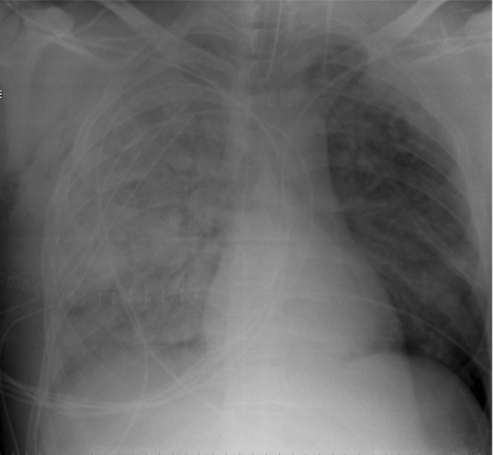
Lung parenchymal injury or lung contusion
Lung contusions and lacerations are predominant injuries.
Often occur without external signs of injury
EdemaEdemaEdema is a condition in which excess serous fluid accumulates in the body cavity or interstitial space of connective tissues. Edema is a symptom observed in several medical conditions. It can be categorized into 2 types, namely, peripheral (in the extremities) and internal (in an organ or body cavity). Edema, hemorrhage, and inflammationInflammationInflammation is a complex set of responses to infection and injury involving leukocytes as the principal cellular mediators in the body’s defense against pathogenic organisms. Inflammation is also seen as a response to tissue injury in the process of wound healing. The 5 cardinal signs of inflammation are pain, heat, redness, swelling, and loss of function. Inflammation of alveolar spaces → ventilationVentilationThe total volume of gas inspired or expired per unit of time, usually measured in liters per minute.Ventilation: Mechanics of Breathing/perfusion mismatch
2 mechanisms: compressionCompressionBlunt Chest Trauma or tearing, and severe displacementDisplacementThe process by which an emotional or behavioral response that is appropriate for one situation appears in another situation for which it is inappropriate.Defense Mechanisms
Alveolar bleeding produces hepatization.
Consolidated area on chest X-rayX-rayPenetrating electromagnetic radiation emitted when the inner orbital electrons of an atom are excited and release radiant energy. X-ray wavelengths range from 1 pm to 10 nm. Hard x-rays are the higher energy, shorter wavelength x-rays. Soft x-rays or grenz rays are less energetic and longer in wavelength. The short wavelength end of the x-ray spectrum overlaps the gamma rays wavelength range. The distinction between gamma rays and x-rays is based on their radiation source.Pulmonary Function Tests (difficult to differentiate from pneumoniaPneumoniaPneumonia or pulmonary inflammation is an acute or chronic inflammation of lung tissue. Causes include infection with bacteria, viruses, or fungi. In more rare cases, pneumonia can also be caused through toxic triggers through inhalation of toxic substances, immunological processes, or in the course of radiotherapy.Pneumonia or atelectasisAtelectasisAtelectasis is the partial or complete collapse of a part of the lung. Atelectasis is almost always a secondary phenomenon from conditions causing bronchial obstruction, external compression, surfactant deficiency, or scarring. Atelectasis)
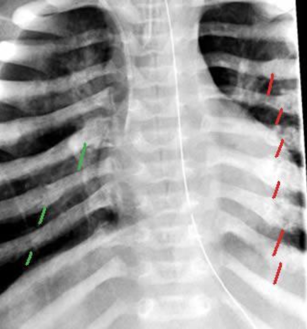
Rib fractureFractureA fracture is a disruption of the cortex of any bone and periosteum and is commonly due to mechanical stress after an injury or accident. Open fractures due to trauma can be a medical emergency. Fractures are frequently associated with automobile accidents, workplace injuries, and trauma.Overview of Bone Fractures
Signifies severe injury due to high-impact forces
May result in flail chestFlail chestFlail chest is a life-threatening traumatic injury that occurs when 3 or more contiguous ribs are fractured in 2 or more different locations. Patients present with chest pain, tachypnea, hypoxia, and paradoxical chest wall movement. Flail Chest
Can indicate child abuseChild abuseChild abuse is an act or failure to act that results in harm to a child’s health or development. The abuse encompasses neglect as well as physical, sexual, and emotional harm. Seen in all subsets of society, child abuse is a cause of significant morbidity and mortality in the pediatric population. Child Abuse when high-energy mechanisms are lacking
Blunt cardiac injury or cardiac contusion
Seen in sport-related blunt trauma to the chest
Affected individuals usually die before reaching the hospital.
Can manifest as arrhythmias, commotio cordisCommotio cordisA sudden cardiac arrhythmia (e.g., ventricular fibrillation) caused by a blunt, non-penetrating impact to the precordial region of chest wall. Commotio cordis often results in sudden death without prompt cardiopulmonary defibrillation.Blunt Chest Trauma, and sudden death
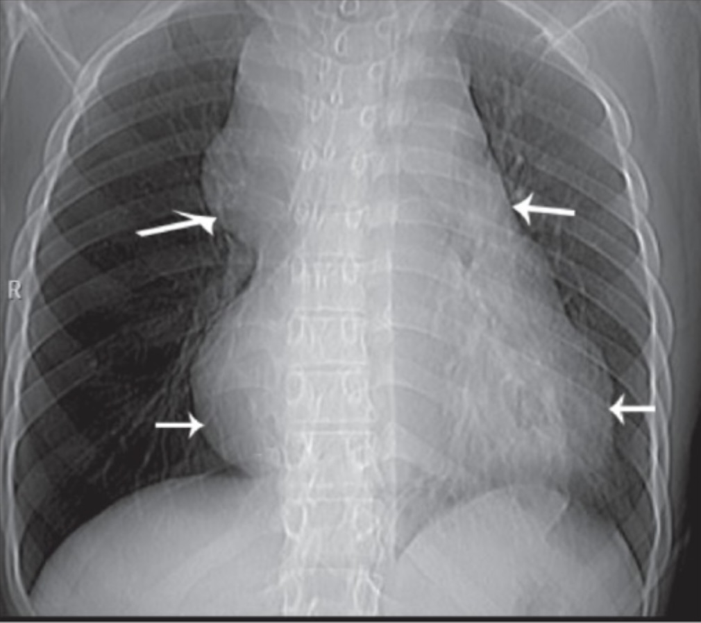
Widening of the mediastinal contour on chest X-rayX-rayPenetrating electromagnetic radiation emitted when the inner orbital electrons of an atom are excited and release radiant energy. X-ray wavelengths range from 1 pm to 10 nm. Hard x-rays are the higher energy, shorter wavelength x-rays. Soft x-rays or grenz rays are less energetic and longer in wavelength. The short wavelength end of the x-ray spectrum overlaps the gamma rays wavelength range. The distinction between gamma rays and x-rays is based on their radiation source.Pulmonary Function Tests
Recognized by pneumomediastinumPneumomediastinumMediastinitis or pneumoperitoneumPneumoperitoneumA condition with trapped gas or air in the peritoneal cavity, usually secondary to perforation of the internal organs such as the lung and the gastrointestinal tract, or to recent surgery. Pneumoperitoneum may be purposely introduced to aid radiological examination.Perforated Viscus in diagnostic imaging (pneumomediastinumPneumomediastinumMediastinitis in 10% of cases of blunt chest traumaBlunt chest traumaBlunt chest trauma is a non-penetrating traumatic injury to the thoracic cavity. Thoracic traumatic injuries are classified according to the mechanism of injury as blunt or penetrating injuries. Different structures can be injured including the chest wall (ribs, sternum), lungs, heart, major blood vessels, and the esophagus.Blunt Chest Trauma)
Diaphragmatic injury
May be accompanied by phrenic nervePhrenic nerveThe motor nerve of the diaphragm. The phrenic nerve fibers originate in the cervical spinal column (mostly C4) and travel through the cervical plexus to the diaphragm.Diaphragm: Anatomy paralysis, liverLiverThe liver is the largest gland in the human body. The liver is found in the superior right quadrant of the abdomen and weighs approximately 1.5 kilograms. Its main functions are detoxification, metabolism, nutrient storage (e.g., iron and vitamins), synthesis of coagulation factors, formation of bile, filtration, and storage of blood. Liver: Anatomy and spleenSpleenThe spleen is the largest lymphoid organ in the body, located in the LUQ of the abdomen, superior to the left kidney and posterior to the stomach at the level of the 9th-11th ribs just below the diaphragm. The spleen is highly vascular and acts as an important blood filter, cleansing the blood of pathogens and damaged erythrocytes. Spleen: Anatomy injury, and respiratory distress
Bowel sounds in the chest may be heard.
Traumatic asphyxiaAsphyxiaA pathological condition caused by lack of oxygen, manifested in impending or actual cessation of life.Drowning
Occurs due to flexibility of the chest wallChest wallThe chest wall consists of skin, fat, muscles, bones, and cartilage. The bony structure of the chest wall is composed of the ribs, sternum, and thoracic vertebrae. The chest wall serves as armor for the vital intrathoracic organs and provides the stability necessary for the movement of the shoulders and arms. Chest Wall: Anatomy
Intrathoracic pressure increases with compressive force from the abdomen against a closed glottisGlottisThe vocal apparatus of the larynx, situated in the middle section of the larynx. Glottis consists of the vocal folds and an opening (rima glottidis) between the folds.Larynx: Anatomy.
PetechiaePetechiaePrimary Skin Lesions appear in the oral mucosaOral mucosaLining of the oral cavity, including mucosa on the gums; the palate; the lip; the cheek; floor of the mouth; and other structures. The mucosa is generally a nonkeratinized stratified squamous epithelium covering muscle, bone, or glands but can show varying degree of keratinization at specific locations.Stomatitis with edemaEdemaEdema is a condition in which excess serous fluid accumulates in the body cavity or interstitial space of connective tissues. Edema is a symptom observed in several medical conditions. It can be categorized into 2 types, namely, peripheral (in the extremities) and internal (in an organ or body cavity). Edema and cyanosisCyanosisA bluish or purplish discoloration of the skin and mucous membranes due to an increase in the amount of deoxygenated hemoglobin in the blood or a structural defect in the hemoglobin molecule.Pulmonary Examination.
Tracheobronchial injury
Due to rapid decelerationDecelerationA decrease in the rate of speed.Blunt Chest Trauma, crush injuries, or force to the chest against a closed glottisGlottisThe vocal apparatus of the larynx, situated in the middle section of the larynx. Glottis consists of the vocal folds and an opening (rima glottidis) between the folds.Larynx: Anatomy
Gunshot woundsGunshot woundsDisruption of structural continuity of the body as a result of the discharge of firearms.Penetrating Chest Injury are an increasingly common cause.
Look at, listen to, and feel the chest wallChest wallThe chest wall consists of skin, fat, muscles, bones, and cartilage. The bony structure of the chest wall is composed of the ribs, sternum, and thoracic vertebrae. The chest wall serves as armor for the vital intrathoracic organs and provides the stability necessary for the movement of the shoulders and arms. Chest Wall: Anatomy for breathing.
In cases of pneumothoraxPneumothoraxA pneumothorax is a life-threatening condition in which air collects in the pleural space, causing partial or full collapse of the lung. A pneumothorax can be traumatic or spontaneous. Patients present with a sudden onset of sharp chest pain, dyspnea, and diminished breath sounds on exam.Pneumothorax, hemothoraxHemothoraxA hemothorax is a collection of blood in the pleural cavity. Hemothorax most commonly occurs due to damage to the intercostal arteries or from a lung laceration following chest trauma. Hemothorax can also occur as a complication of disease, or hemothorax may be spontaneous or iatrogenic. Hemothorax, or flail chestFlail chestFlail chest is a life-threatening traumatic injury that occurs when 3 or more contiguous ribs are fractured in 2 or more different locations. Patients present with chest pain, tachypnea, hypoxia, and paradoxical chest wall movement. Flail Chest, breathing may be asymmetric.
CirculationCirculationThe movement of the blood as it is pumped through the cardiovascular system.ABCDE Assessment:
DisabilityDisabilityDetermination of the degree of a physical, mental, or emotional handicap. The diagnosis is applied to legal qualification for benefits and income under disability insurance and to eligibility for social security and workman’s compensation benefits.ABCDE Assessment:
Assess the level of alertness.
GCSGCSA scale that assesses the response to stimuli in patients with craniocerebral injuries. The parameters are eye opening, motor response, and verbal response.Coma < 8 is an indication for emergent intubationIntubationPeritonsillar Abscess.
Exposure:
Complete; head-to-toe examination for other injuries
As a shortcut, when the affected individual speaks, it is an indication of:
Sufficient circulationCirculationThe movement of the blood as it is pumped through the cardiovascular system.ABCDE Assessment and perfusion of the brainBrainThe part of central nervous system that is contained within the skull (cranium). Arising from the neural tube, the embryonic brain is comprised of three major parts including prosencephalon (the forebrain); mesencephalon (the midbrain); and rhombencephalon (the hindbrain). The developed brain consists of cerebrum; cerebellum; and other structures in the brain stem.Nervous System: Anatomy, Structure, and Classification
Helpful in assessing mental status
Table: Life-threatening injuries to be considered during the primary survey of thoracic trauma
Massive hemothoraxHemothoraxA hemothorax is a collection of blood in the pleural cavity. Hemothorax most commonly occurs due to damage to the intercostal arteries or from a lung laceration following chest trauma. Hemothorax can also occur as a complication of disease, or hemothorax may be spontaneous or iatrogenic. Hemothorax
Cardiac contusion
Cardiac tamponadeTamponadePericardial effusion, usually of rapid onset, exceeding ventricular filling pressures and causing collapse of the heart with a markedly reduced cardiac output.Pericarditis
Abnormal respiratory rateRespiratory rateThe number of times an organism breathes with the lungs (respiration) per unit time, usually per minute.Pulmonary Examination
Paradoxical chest movements (e.g., flail chestFlail chestFlail chest is a life-threatening traumatic injury that occurs when 3 or more contiguous ribs are fractured in 2 or more different locations. Patients present with chest pain, tachypnea, hypoxia, and paradoxical chest wall movement. Flail Chest)
Image: “Fractures of ribs in an infant” by National Institute of Health. License: Public Domain
Pulmonary contusion on a chest X-ray: See the diffuse “spotting” of the right hemithorax.
Image: “Diffuse bilateral lung infiltrates” by Centre de Santé et de Services Sociaux de Trois-Rivières, 1991 Boul, du Carmel, Trois-Rivières, QC G8Z 3R9, Canada. License: CC BY 2.0
Hemothorax on a chest X-ray: See the “white-out” phenomenon produced by the blood trapped in the pleural space.
Image: “Chest X-ray shows massive right hemothorax” by Cardiac Surgeon, Leeds General Infirmary, Great George Street, Leeds LS1 3EX, UK. License: CC BY 2.0
A widened mediastinum in a chest X-ray seen in cases of aortic disruption
Image: “Frontal chest radiograph shows mediastinal widening” by Department of Radiodiagnosis, R.N.T. Medical College and Associated Group of Hospitals, Udaipur (Rajasthan)-313 004, India. License: CC BY 2.0
Workup
Diagnostic imaging:
Ultrasound (eFAST) in cases of hemodynamic instability
Chest X-rayX-rayPenetrating electromagnetic radiation emitted when the inner orbital electrons of an atom are excited and release radiant energy. X-ray wavelengths range from 1 pm to 10 nm. Hard x-rays are the higher energy, shorter wavelength x-rays. Soft x-rays or grenz rays are less energetic and longer in wavelength. The short wavelength end of the x-ray spectrum overlaps the gamma rays wavelength range. The distinction between gamma rays and x-rays is based on their radiation source.Pulmonary Function Tests: to determine rib fracturesRib fracturesFractures of any of the ribs.Flail Chest, areas of consolidationConsolidationPulmonary Function Tests (lung contusion), pneumothoraxPneumothoraxA pneumothorax is a life-threatening condition in which air collects in the pleural space, causing partial or full collapse of the lung. A pneumothorax can be traumatic or spontaneous. Patients present with a sudden onset of sharp chest pain, dyspnea, and diminished breath sounds on exam.Pneumothorax, and hemothoraxHemothoraxA hemothorax is a collection of blood in the pleural cavity. Hemothorax most commonly occurs due to damage to the intercostal arteries or from a lung laceration following chest trauma. Hemothorax can also occur as a complication of disease, or hemothorax may be spontaneous or iatrogenic. Hemothorax
CT: to determine aortic disruption and hemothoraxHemothoraxA hemothorax is a collection of blood in the pleural cavity. Hemothorax most commonly occurs due to damage to the intercostal arteries or from a lung laceration following chest trauma. Hemothorax can also occur as a complication of disease, or hemothorax may be spontaneous or iatrogenic. Hemothorax
BronchoscopyBronchoscopyEndoscopic examination, therapy or surgery of the bronchi.Laryngomalacia and Tracheomalacia and laryngoscopy: to identify lack of continuity in the upper airways and bronchial treeBronchial treeThe collective term “bronchial tree” refers to the bronchi and all of their subsequent branches. The bronchi are the airways of the lower respiratory tract. At the level of the 3rd or 4th thoracic vertebra, the trachea bifurcates into the left and right main bronchi. Both of these bronchi continue to divide into secondary or lobar bronchi that bifurcate further and further.Bronchial Tree: Anatomy
Esophagoscopy: to determine lack of continuity of the esophagusEsophagusThe esophagus is a muscular tube-shaped organ of around 25 centimeters in length that connects the pharynx to the stomach. The organ extends from approximately the 6th cervical vertebra to the 11th thoracic vertebra and can be divided grossly into 3 parts: the cervical part, the thoracic part, and the abdominal part. Esophagus: Anatomy
Laboratory assessments: troponin levels to rule out blunt cardiac injury
ECGECGAn electrocardiogram (ECG) is a graphic representation of the electrical activity of the heart plotted against time. Adhesive electrodes are affixed to the skin surface allowing measurement of cardiac impulses from many angles. The ECG provides 3-dimensional information about the conduction system of the heart, the myocardium, and other cardiac structures. Electrocardiogram (ECG): to check for arrhythmias
Management depends on the injury (rapid initiation of high-quality CPRCPRThe artificial substitution of heart and lung action as indicated for heart arrest resulting from electric shock, drowning, respiratory arrest, or other causes. The two major components of cardiopulmonary resuscitation are artificial ventilation and closed-chest cardiac massage.Cardiac Arrest if the individual fails the airwayAirwayABCDE Assessment, breathing, circulationCirculationThe movement of the blood as it is pumped through the cardiovascular system.ABCDE Assessment (ABC) assessment).
Positive pressure ventilationPositive pressure ventilationApplication of positive pressure to the inspiratory phase when the patient has an artificial airway in place and is connected to a ventilator.Flail Chest, if necessary
Maintain blood pressure/perfusion:
IsotonicIsotonicSolutions having the same osmotic pressure as blood serum, or another solution with which they are compared.Renal Sodium and Water Regulation crystalloids or blood transfusion, if necessary
Flail chestFlail chestFlail chest is a life-threatening traumatic injury that occurs when 3 or more contiguous ribs are fractured in 2 or more different locations. Patients present with chest pain, tachypnea, hypoxia, and paradoxical chest wall movement. Flail Chest:
Surgical fixation of chest wallChest wallThe chest wall consists of skin, fat, muscles, bones, and cartilage. The bony structure of the chest wall is composed of the ribs, sternum, and thoracic vertebrae. The chest wall serves as armor for the vital intrathoracic organs and provides the stability necessary for the movement of the shoulders and arms. Chest Wall: Anatomy when necessary
HemothoraxHemothoraxA hemothorax is a collection of blood in the pleural cavity. Hemothorax most commonly occurs due to damage to the intercostal arteries or from a lung laceration following chest trauma. Hemothorax can also occur as a complication of disease, or hemothorax may be spontaneous or iatrogenic. Hemothorax: tube thoracostomyTube ThoracostomySurgical procedure involving the creation of an opening (stoma) into the chest cavity for drainage; used in the treatment of pleural effusion; pneumothorax; hemothorax; and empyema.Thoracic Surgery or thoracotomyThoracotomySurgical incision into the chest wall.Thoracic Surgery in cases of severe bleeding
Gutiérrez, C.E. (2016). Pediatric trauma. In J. E. Tintinalli, J.S. Stapczynski, O.J. Ma, D.M. Yealy, G.D. Meckler, D.M. Cline (Eds.), Tintinalli’s Emergency Medicine: A Comprehensive Study Guide, 8e. New York, NY: McGraw-Hill Education. accessmedicine.mhmedical.com/content.aspx?aid=1121492666
Roskind, C.G., Pryor, H.I., Klein, B.L. (2020). Acute care of multiple trauma. In R.M. Kliegman MD, J.W. St Geme, N.J. Blum, S.S. Shah, MSCE, R.C. Tasker, K.M. Wilson (Eds.), Nelson Textbook of Pediatrics (pp. 54-554.e1). https://www.clinicalkey.es/#!/content/3-s2.0-B9780323529501000821