The chest wall consists of skin Skin The skin, also referred to as the integumentary system, is the largest organ of the body. The skin is primarily composed of the epidermis (outer layer) and dermis (deep layer). The epidermis is primarily composed of keratinocytes that undergo rapid turnover, while the dermis contains dense layers of connective tissue. Skin: Structure and Functions, fat, muscles, bones, and cartilage Cartilage Cartilage is a type of connective tissue derived from embryonic mesenchyme that is responsible for structural support, resilience, and the smoothness of physical actions. Perichondrium (connective tissue membrane surrounding cartilage) compensates for the absence of vasculature in cartilage by providing nutrition and support. Cartilage: Histology. The bony structure of the chest wall is composed of the ribs, sternum, and thoracic vertebrae Thoracic vertebrae A group of twelve vertebrae connected to the ribs that support the upper trunk region. Vertebral Column: Anatomy. The chest wall serves as armor for the vital intrathoracic organs and provides the stability necessary for the movement of the shoulders and arms. The interconnections between the bones, cartilage Cartilage Cartilage is a type of connective tissue derived from embryonic mesenchyme that is responsible for structural support, resilience, and the smoothness of physical actions. Perichondrium (connective tissue membrane surrounding cartilage) compensates for the absence of vasculature in cartilage by providing nutrition and support. Cartilage: Histology, and muscles allow for the rhythmic expansion and reduction of the chest wall during breathing, which facilitates changes in intrathoracic pressure to allow expansion of the lungs Lungs Lungs are the main organs of the respiratory system. Lungs are paired viscera located in the thoracic cavity and are composed of spongy tissue. The primary function of the lungs is to oxygenate blood and eliminate CO2. Lungs: Anatomy during inspiration Inspiration Ventilation: Mechanics of Breathing. The extrinsic muscles have 2 bony attachments; the intrinsic muscles only attach to the thoracic skeleton.
Last updated: Dec 15, 2025
The thoracic skeleton consists of 12 pairs of ribs.
Classification:
Parts of each rib:
Intercostal spaces:
The spaces between the ribs are the intercostal spaces:
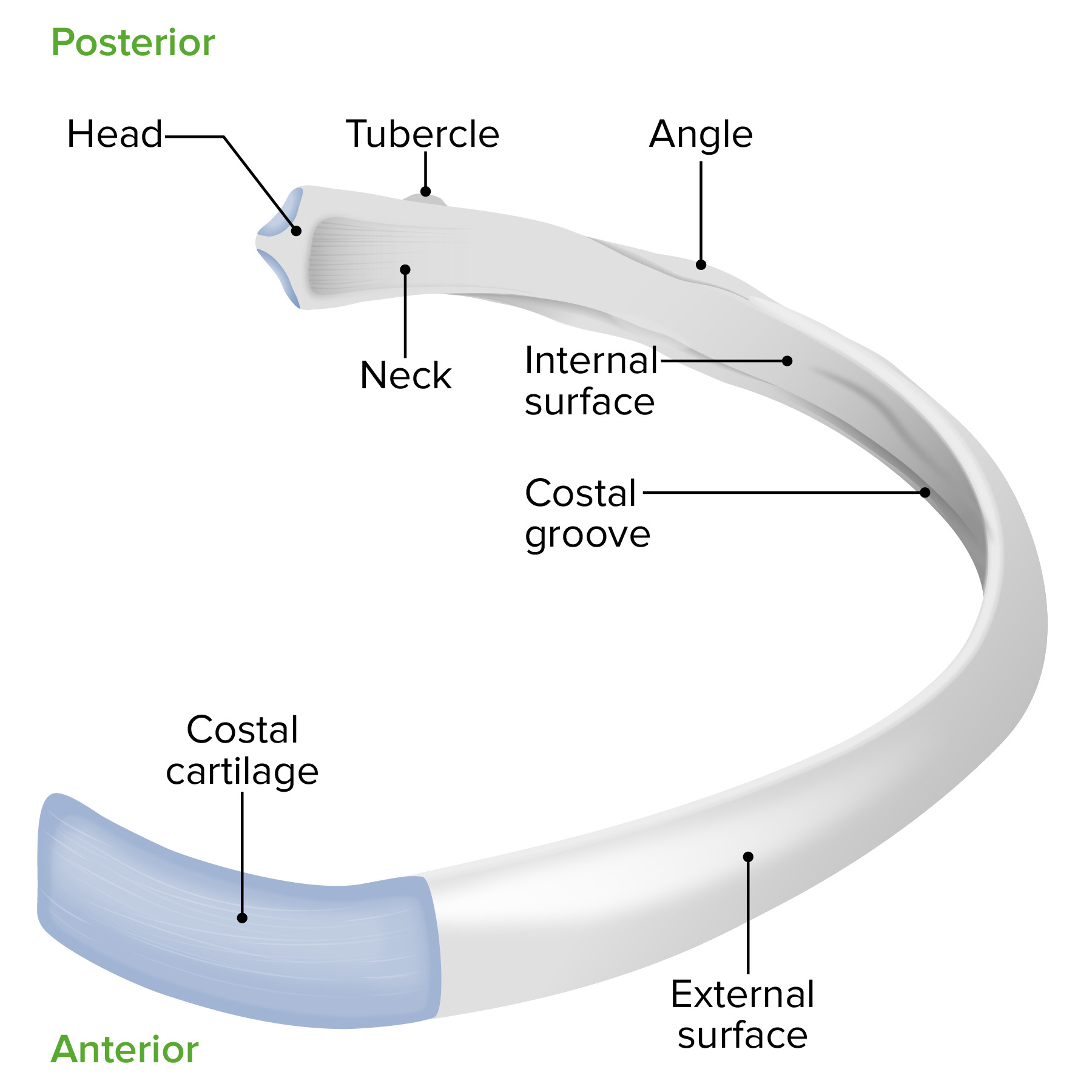
Anatomy of a rib
Image by Lecturio.Consists of the following parts:
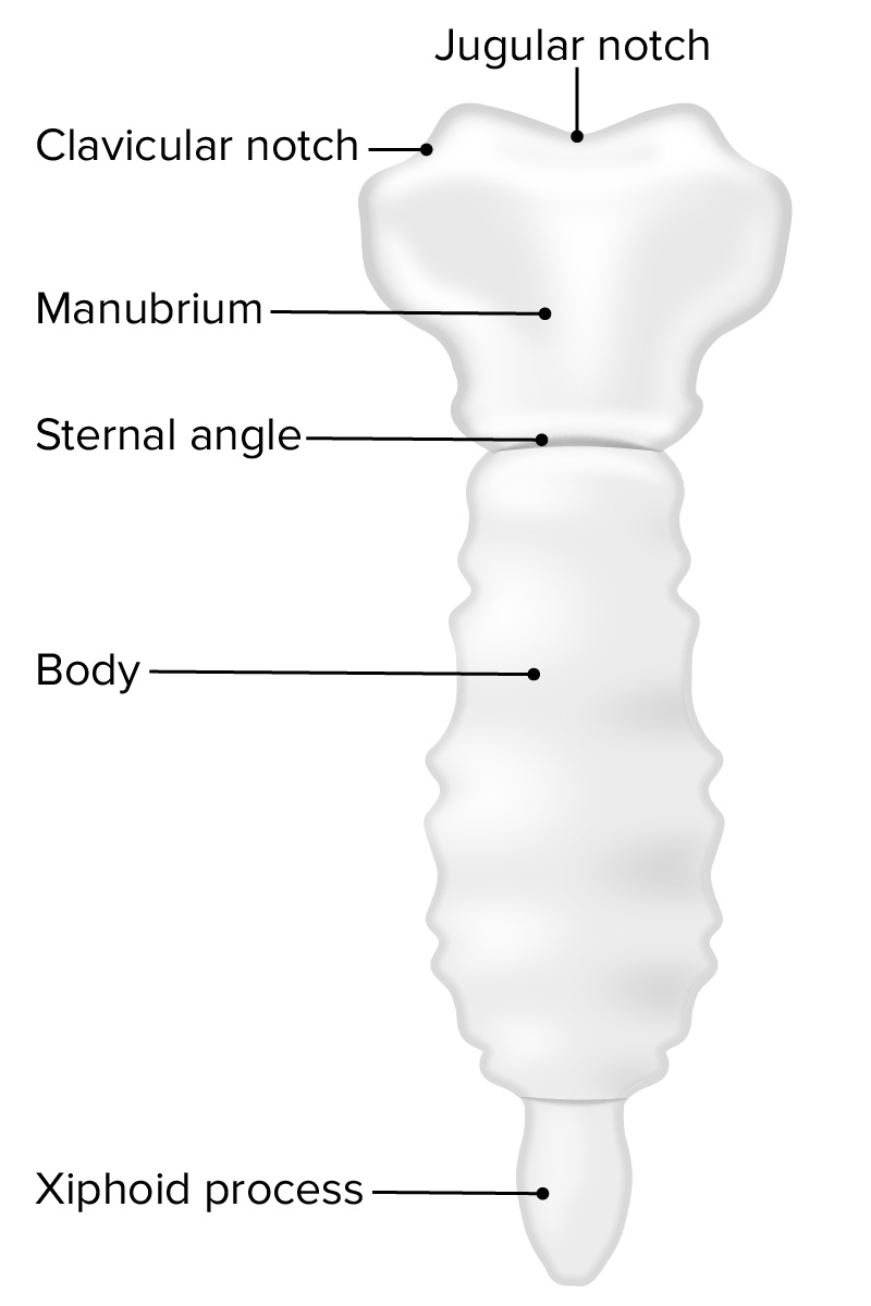
Anatomy of the sternum
Image by Lecturio.Intrinsic muscles attach only to the thorax. The intrinsic muscles receive blood supply from the intercostal arteries Arteries Arteries are tubular collections of cells that transport oxygenated blood and nutrients from the heart to the tissues of the body. The blood passes through the arteries in order of decreasing luminal diameter, starting in the largest artery (the aorta) and ending in the small arterioles. Arteries are classified into 3 types: large elastic arteries, medium muscular arteries, and small arteries and arterioles. Arteries: Histology and are innervated by the intercostal nerves.
External intercostal muscles:
Internal intercostal muscles:
Innermost intercostal muscles:
Subcostal muscles:
Transversus thoracis muscles:
Levatores costarum muscles:
Serratus posterior muscles:
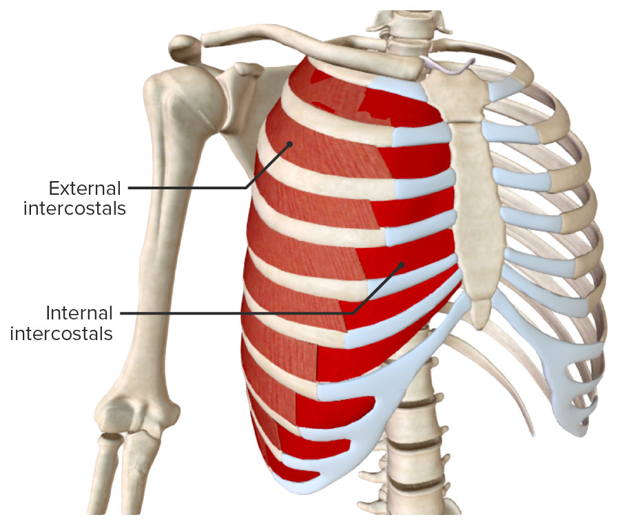
External and internal intercostal muscles
Image by BioDigital, edited by Lecturio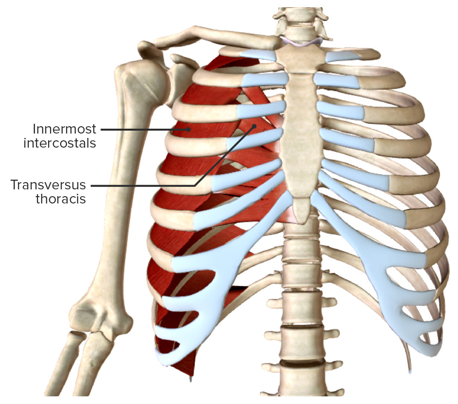
Innermost intercostals and transversus thoracis muscles
Image by BioDigital, edited by Lecturio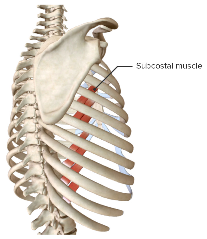
Subcostal muscle
Image by BioDigital, edited by Lecturio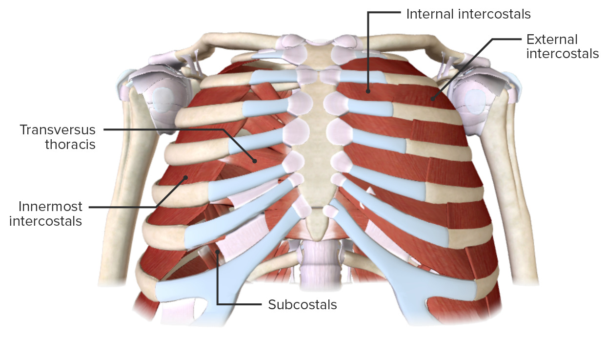
Intrinsic muscles of the chest wall
Image by BioDigital, edited by Lecturio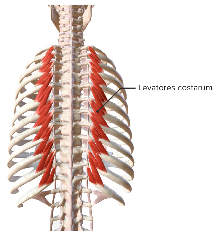
Levatores costarum muscle
Image by BioDigital, edited by Lecturio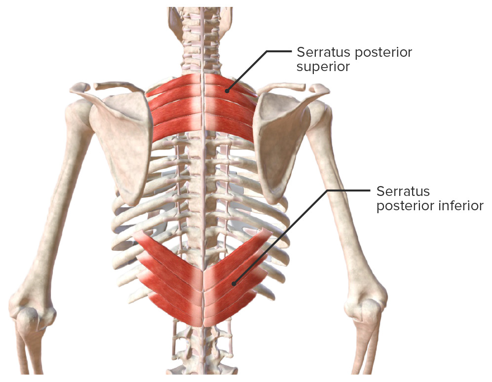
Serratus posterior muscles (superior and inferior)
Image by BioDigital, edited by LecturioIn contrast to the intrinsic muscles, the extrinsic muscles attach to the thorax and other areas of the body.
Pectoralis major muscle:
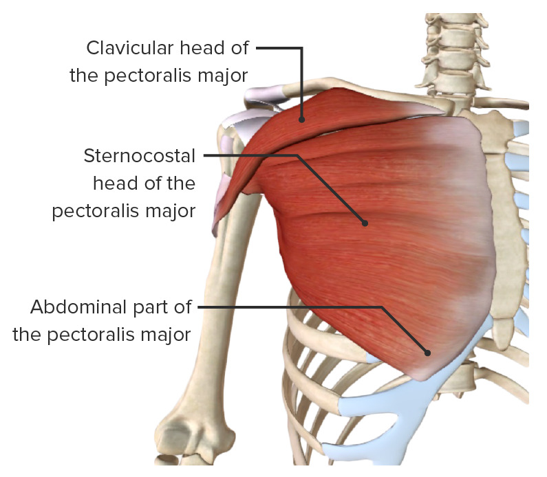
Pectoralis major muscle
Image by BioDigital, edited by LecturioPectoralis minor muscle:
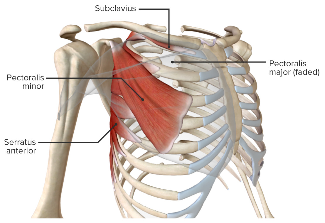
Extrinsic muscles of the chest wall
Image by Lecturio.Serratus anterior muscle:
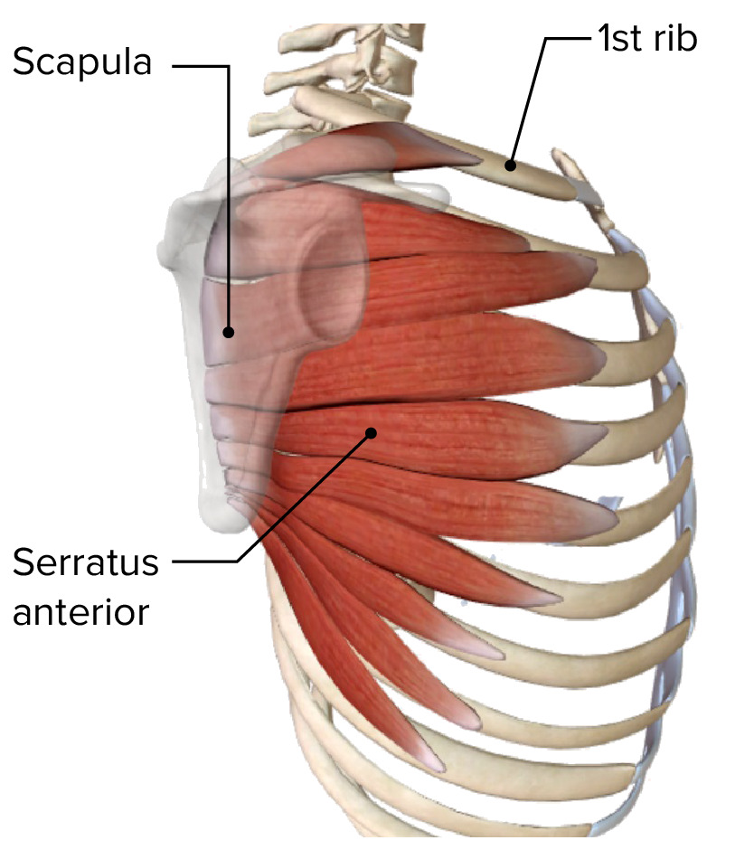
Serratus anterior muscle
Image by BioDigital, edited by LecturioSubclavius Subclavius Muscles of the Neck: Anatomy muscle:
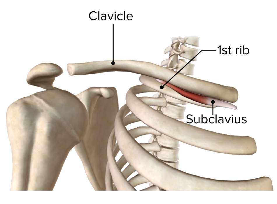
Subclavius muscle
Image by BioDigital, edited by Lecturio