The neck Neck The part of a human or animal body connecting the head to the rest of the body. Peritonsillar Abscess is considered to be quadrangular. This shape is the basis to study various components of the neck Neck The part of a human or animal body connecting the head to the rest of the body. Peritonsillar Abscess and their relations. The boundaries of the quadrangular shape include the mandible Mandible The largest and strongest bone of the face constituting the lower jaw. It supports the lower teeth. Jaw and Temporomandibular Joint: Anatomy, upper border of the clavicle Clavicle A bone on the ventral side of the shoulder girdle, which in humans is commonly called the collar bone. Clavicle Fracture, midline of the neck Neck The part of a human or animal body connecting the head to the rest of the body. Peritonsillar Abscess, and anterior margin of the trapezius. The quadrangular shape is divided into an anterior and posterior triangle by the sternocleidomastoid Sternocleidomastoid Muscles of the Neck: Anatomy muscle (SCM). The anterior and posterior triangles are the 2 primary subdivisions and are delineated by easily recognized anatomic structures. Each triangle houses muscles, nerves, vasculature, lymphatics, and adipose tissue Adipose tissue Adipose tissue is a specialized type of connective tissue that has both structural and highly complex metabolic functions, including energy storage, glucose homeostasis, and a multitude of endocrine capabilities. There are three types of adipose tissue, white adipose tissue, brown adipose tissue, and beige or "brite" adipose tissue, which is a transitional form. Adipose Tissue: Histology.
Last updated: Dec 15, 2025
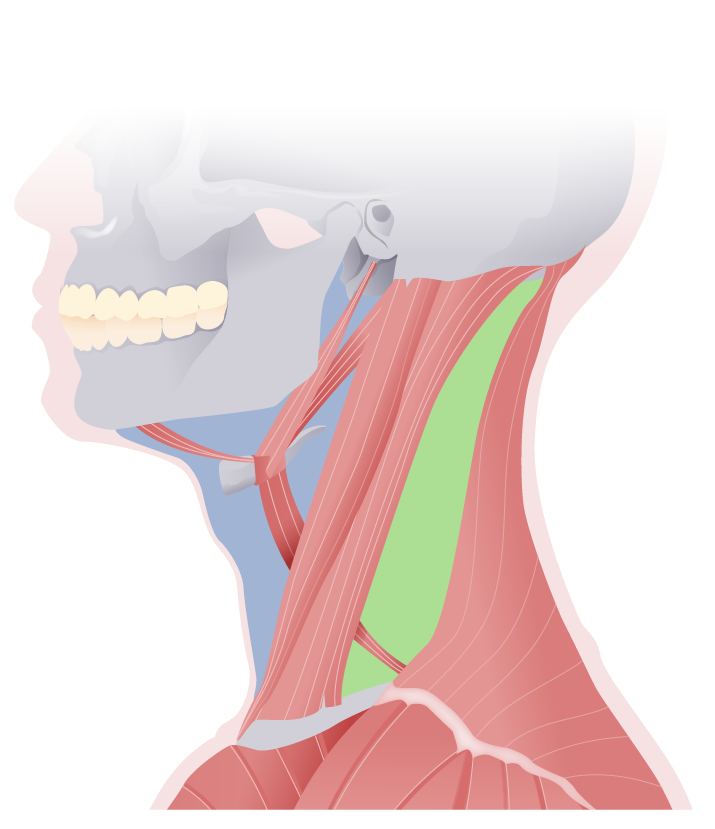
Lateral view of the neck:
featuring the anterior (blue) and posterior (green) triangles of the neck, separated by the sternocleidomastoid muscle
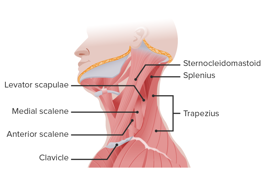
Primary muscles of the neck
Image by Lecturio.Boundaries:
Contents:
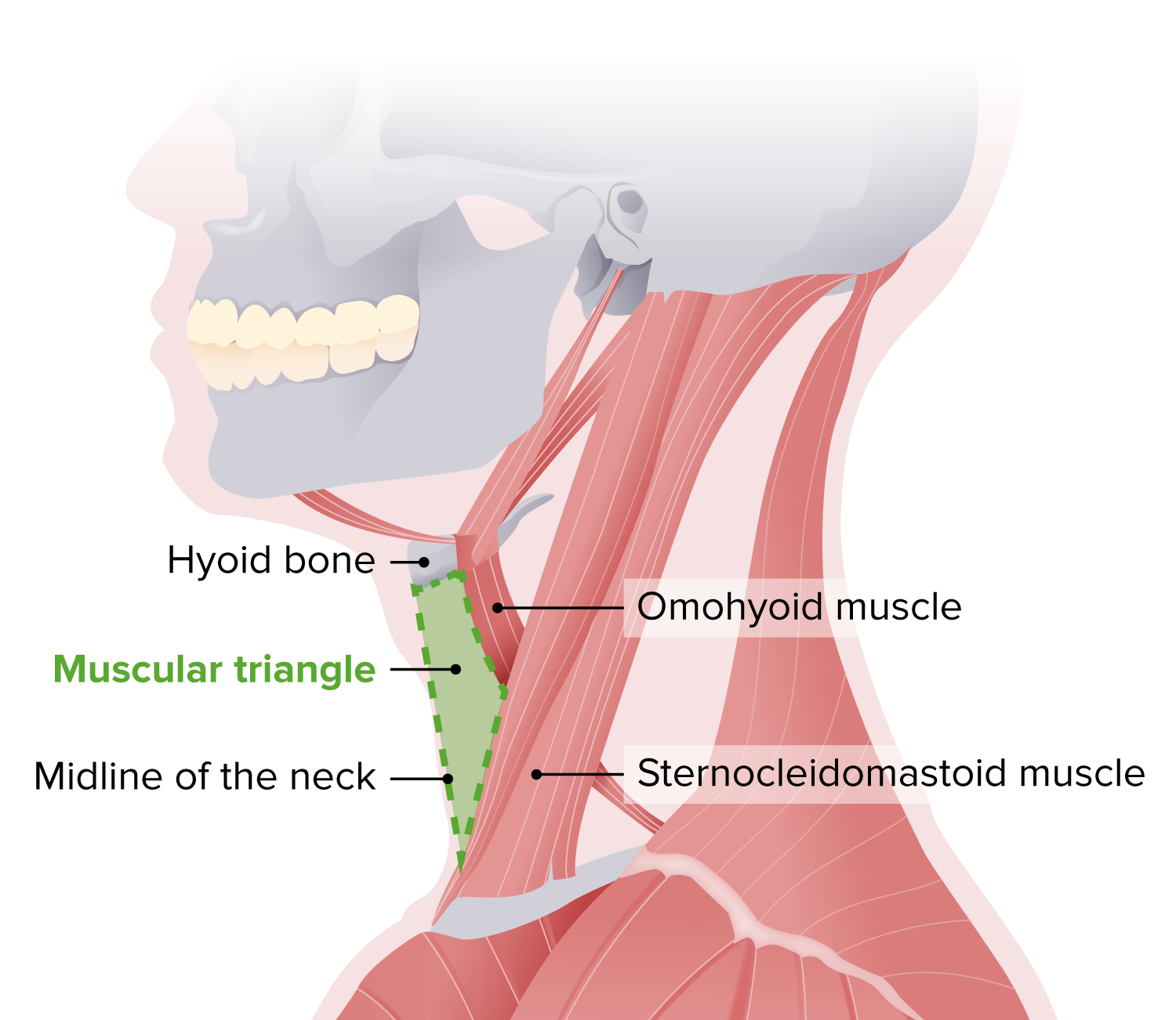
Muscular triangle of the neck
Image by Lecturio.Boundaries:
Contents:
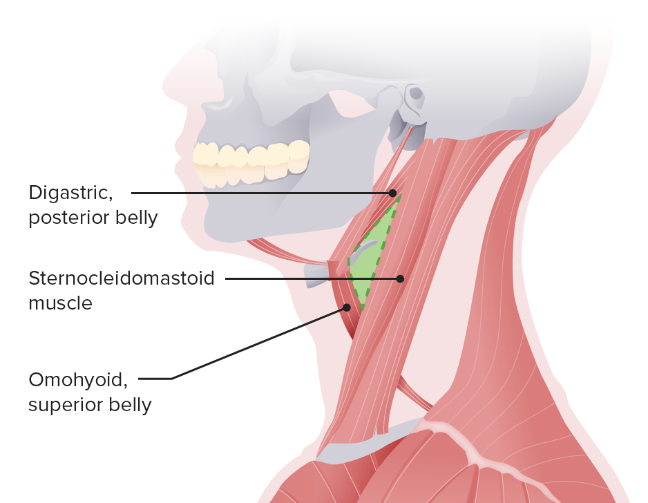
Carotid triangle
Image by Lecturio.Boundaries:
Contents:
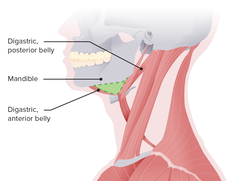
Submandibular triangle
Image by Lecturio.Boundaries:
Contents:
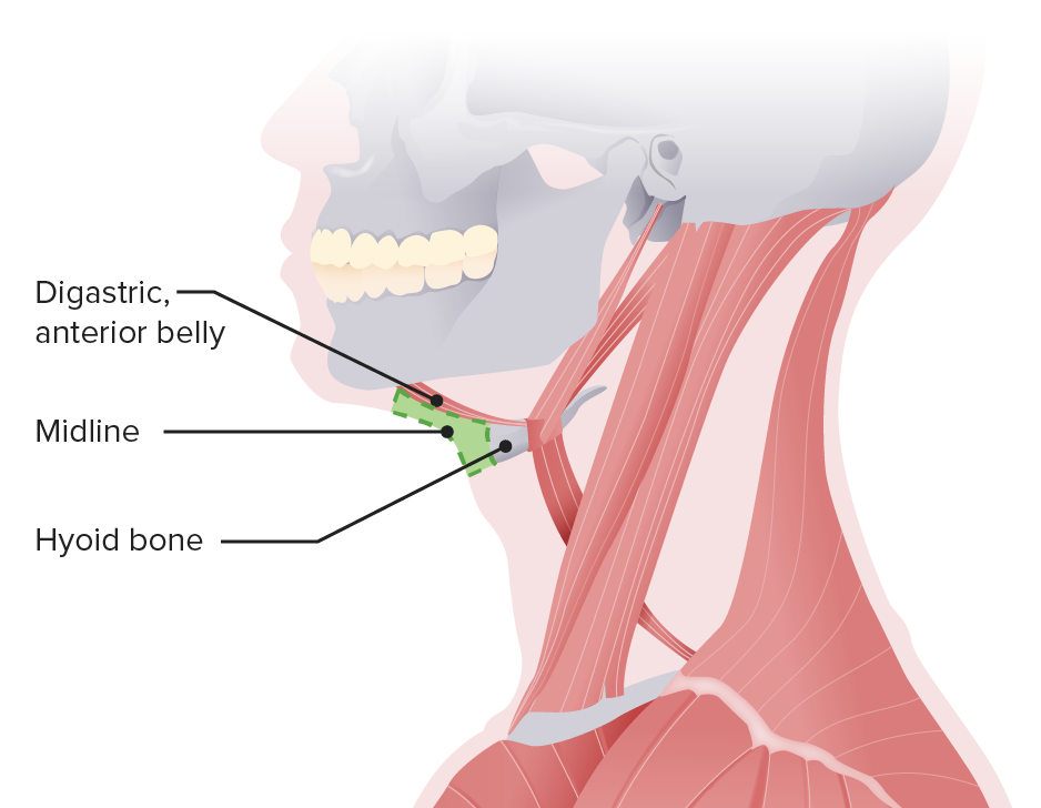
Submental triangle
Image by Lecturio.Boundaries:
Contents:
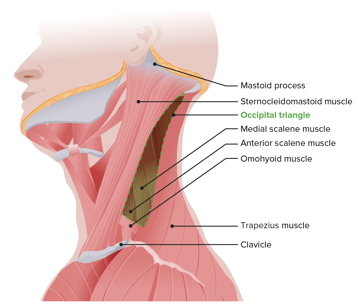
Schematic of the occipital triangle
Image by Lecturio.The supraclavicular triangle is also called the omoclavicular or subclavian triangle, and it is smaller with the arm Arm The arm, or “upper arm” in common usage, is the region of the upper limb that extends from the shoulder to the elbow joint and connects inferiorly to the forearm through the cubital fossa. It is divided into 2 fascial compartments (anterior and posterior). Arm: Anatomy raised and bigger with the arm Arm The arm, or “upper arm” in common usage, is the region of the upper limb that extends from the shoulder to the elbow joint and connects inferiorly to the forearm through the cubital fossa. It is divided into 2 fascial compartments (anterior and posterior). Arm: Anatomy/ clavicle Clavicle A bone on the ventral side of the shoulder girdle, which in humans is commonly called the collar bone. Clavicle Fracture depressed.
Boundaries:
Contents:
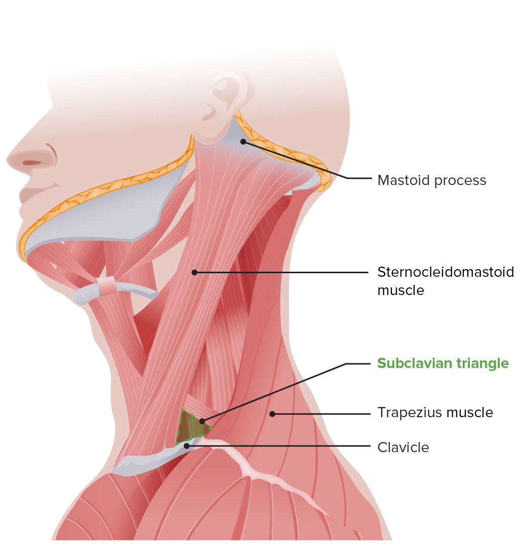
Schematic of the supraclavicular triangle
Image by Lecturio.