Achieve Mastery of Medical Concepts

Study for medical school and boards with Lecturio

Pleura: Anatomy

The pleura is a serous membrane that lines the walls of the thoracic cavity and the surface of the lungs Lungs Lungs are the main organs of the respiratory system. Lungs are paired viscera located in the thoracic cavity and are composed of spongy tissue. The primary function of the lungs is to oxygenate blood and eliminate CO2. Lungs: Anatomy. This structure of mesodermal origin covers both lungs Lungs Lungs are the main organs of the respiratory system. Lungs are paired viscera located in the thoracic cavity and are composed of spongy tissue. The primary function of the lungs is to oxygenate blood and eliminate CO2. Lungs: Anatomy, the mediastinum Mediastinum The mediastinum is the thoracic area between the 2 pleural cavities. The mediastinum contains vital structures of the circulatory, respiratory, digestive, and nervous systems including the heart and esophagus, and major thoracic vessels. Mediastinum and Great Vessels: Anatomy, the thoracic surface of the diaphragm Diaphragm The diaphragm is a large, dome-shaped muscle that separates the thoracic cavity from the abdominal cavity. The diaphragm consists of muscle fibers and a large central tendon, which is divided into right and left parts. As the primary muscle of inspiration, the diaphragm contributes 75% of the total inspiratory muscle force. Diaphragm: Anatomy, and the inner part of the thoracic cage. The pleura is divided into a visceral pleura and parietal pleura Parietal pleura Pleuritis. Between both layers, there is a well-lubricated potential space called the pleural cavity, which eases the respiratory movements of the lungs Lungs Lungs are the main organs of the respiratory system. Lungs are paired viscera located in the thoracic cavity and are composed of spongy tissue. The primary function of the lungs is to oxygenate blood and eliminate CO2. Lungs: Anatomy and helps avoid friction.

Last updated: Jan 17, 2024

Editorial responsibility: Stanley Oiseth, Lindsay Jones, Evelin Maza

Gross Anatomy

The pleura is a double-layered serous membrane that lines the walls of the thoracic cavity and the surface of the lungs Lungs Lungs are the main organs of the respiratory system. Lungs are paired viscera located in the thoracic cavity and are composed of spongy tissue. The primary function of the lungs is to oxygenate blood and eliminate CO2. Lungs: Anatomy. Thus, it extends virtually as far and wide as the thoracic cavity.

Boundaries: 

  • Superior: root of neck Neck The part of a human or animal body connecting the head to the rest of the body. Peritonsillar Abscess 2–3 cm above rib 1
  • Inferior: thoracic surface of the diaphragm Diaphragm The diaphragm is a large, dome-shaped muscle that separates the thoracic cavity from the abdominal cavity. The diaphragm consists of muscle fibers and a large central tendon, which is divided into right and left parts. As the primary muscle of inspiration, the diaphragm contributes 75% of the total inspiratory muscle force. Diaphragm: Anatomy
  • Medial: mediastinum Mediastinum The mediastinum is the thoracic area between the 2 pleural cavities. The mediastinum contains vital structures of the circulatory, respiratory, digestive, and nervous systems including the heart and esophagus, and major thoracic vessels. Mediastinum and Great Vessels: Anatomy
  • Lateral: inner surface of the rib cage Rib cage The bony thoracic enclosure consisting of the vertebral column; the ribs; the sternum; and the costal cartilage. Chest Wall: Anatomy and intercostal muscles Intercostal Muscles Respiratory muscles that arise from the lower border of one rib and insert into the upper border of the adjoining rib, and contract during inspiration or respiration. Chest Wall: Anatomy
Boundaries and parts of the pleura within the thoracic cavity

Boundaries and parts of the pleura within the thoracic cavity

Image by Lecturio. License: CC BY-NC-SA 4.0

Each lung is enclosed in a serous pleural sac that consists of 2 continuous membranes of visceral and parietal pleura Parietal pleura Pleuritis.

  • Parietal pleura Parietal pleura Pleuritis:
    • Lines the inner surface of the thoracic cavity
    • Separated from the thoracic wall by the endothoracic fascia Fascia Layers of connective tissue of variable thickness. The superficial fascia is found immediately below the skin; the deep fascia invests muscles, nerves, and other organs. Cellulitis
    • Parts are classified according to the adjacent structures:
      • Costal pleura
      • Diaphragmatic pleura
      • Mediastinal pleura: forms a sleeve-like membranous tube called the root of the lung and covers the heart and great vessels of the heart
      • Cervical pleura: covered by the suprapleural membrane, a dome-shaped fascia Fascia Layers of connective tissue of variable thickness. The superficial fascia is found immediately below the skin; the deep fascia invests muscles, nerves, and other organs. Cellulitis attached to the 1st rib and C7 that serves as a site of insertion for some of the deep muscles of the neck Neck The part of a human or animal body connecting the head to the rest of the body. Peritonsillar Abscess
  • Visceral pleura:
    • Lines the outer surface of the lungs Lungs Lungs are the main organs of the respiratory system. Lungs are paired viscera located in the thoracic cavity and are composed of spongy tissue. The primary function of the lungs is to oxygenate blood and eliminate CO2. Lungs: Anatomy
    • Covers lung fissures
    • Not sensitive to pain Pain An unpleasant sensation induced by noxious stimuli which are detected by nerve endings of nociceptive neurons. Pain: Types and Pathways
    • Continues with parietal pleura Parietal pleura Pleuritis at the hilum Hilum Lungs: Anatomy of each lung
  • Recesses: named according to the points of reflection of the pleura
    • Costodiaphragmatic recess:
      • The largest recess
      • Found between the rib cage Rib cage The bony thoracic enclosure consisting of the vertebral column; the ribs; the sternum; and the costal cartilage. Chest Wall: Anatomy and the diaphragmatic portion of the pleura in each side of the thoracic cavity  
    • Costomedial recess:
      • Found anteriorly between the rib cage Rib cage The bony thoracic enclosure consisting of the vertebral column; the ribs; the sternum; and the costal cartilage. Chest Wall: Anatomy and the mediastinum Mediastinum The mediastinum is the thoracic area between the 2 pleural cavities. The mediastinum contains vital structures of the circulatory, respiratory, digestive, and nervous systems including the heart and esophagus, and major thoracic vessels. Mediastinum and Great Vessels: Anatomy in each side of the thoracic cavity
      • The left recess is larger than the right due to the cardiac notch Cardiac notch Lungs: Anatomy of the left lung.
    • Vertebromediastinal recess:
Layers of thoracic wall - intercostal neurovascular bundle

Layers of the thoracic wall:
Note the double layer of pleura and the pleural cavity, separated from the rib cage by the endothoracic fascia.

Image by Lecturio. License: CC BY-NC-SA 4.0

Pleural cavity:

  • The potential space between the visceral pleura and parietal pleura Parietal pleura Pleuritis (virtually nonexistent/undetectable)
  • Contains a small amount (0.1–0.2 mL/kg) of serous fluid that helps avoid friction between both pleurae
  • The surface tension Surface tension The force acting on the surface of a liquid, tending to minimize the area of the surface. Acute Respiratory Distress Syndrome (ARDS) of the fluid keeps the lungs Lungs Lungs are the main organs of the respiratory system. Lungs are paired viscera located in the thoracic cavity and are composed of spongy tissue. The primary function of the lungs is to oxygenate blood and eliminate CO2. Lungs: Anatomy expanded and in contact with the thoracic wall through the double layer of pleura.

Related videos

Neurovasculature

Table: Neurovasculature of the pleura
Irrigation Innervation
Parietal pleura Parietal pleura Pleuritis Costal portion is supplied by:
  • Branches of the intercostal arteries Arteries Arteries are tubular collections of cells that transport oxygenated blood and nutrients from the heart to the tissues of the body. The blood passes through the arteries in order of decreasing luminal diameter, starting in the largest artery (the aorta) and ending in the small arterioles. Arteries are classified into 3 types: large elastic arteries, medium muscular arteries, and small arteries and arterioles. Arteries: Histology
  • Branches of the internal thoracic arteries Arteries Arteries are tubular collections of cells that transport oxygenated blood and nutrients from the heart to the tissues of the body. The blood passes through the arteries in order of decreasing luminal diameter, starting in the largest artery (the aorta) and ending in the small arterioles. Arteries are classified into 3 types: large elastic arteries, medium muscular arteries, and small arteries and arterioles. Arteries: Histology
Mediastinal portion is supplied by:
  • Bronchial arteries Arteries Arteries are tubular collections of cells that transport oxygenated blood and nutrients from the heart to the tissues of the body. The blood passes through the arteries in order of decreasing luminal diameter, starting in the largest artery (the aorta) and ending in the small arterioles. Arteries are classified into 3 types: large elastic arteries, medium muscular arteries, and small arteries and arterioles. Arteries: Histology
  • Upper diaphragmatic arteries Arteries Arteries are tubular collections of cells that transport oxygenated blood and nutrients from the heart to the tissues of the body. The blood passes through the arteries in order of decreasing luminal diameter, starting in the largest artery (the aorta) and ending in the small arterioles. Arteries are classified into 3 types: large elastic arteries, medium muscular arteries, and small arteries and arterioles. Arteries: Histology
  • Internal thoracic arteries Arteries Arteries are tubular collections of cells that transport oxygenated blood and nutrients from the heart to the tissues of the body. The blood passes through the arteries in order of decreasing luminal diameter, starting in the largest artery (the aorta) and ending in the small arterioles. Arteries are classified into 3 types: large elastic arteries, medium muscular arteries, and small arteries and arterioles. Arteries: Histology
  • Mediastinal arteries Arteries Arteries are tubular collections of cells that transport oxygenated blood and nutrients from the heart to the tissues of the body. The blood passes through the arteries in order of decreasing luminal diameter, starting in the largest artery (the aorta) and ending in the small arterioles. Arteries are classified into 3 types: large elastic arteries, medium muscular arteries, and small arteries and arterioles. Arteries: Histology
Cervical portion Cervical portion Trachea: Anatomy is supplied by: branches of the subclavian arteries Arteries Arteries are tubular collections of cells that transport oxygenated blood and nutrients from the heart to the tissues of the body. The blood passes through the arteries in order of decreasing luminal diameter, starting in the largest artery (the aorta) and ending in the small arterioles. Arteries are classified into 3 types: large elastic arteries, medium muscular arteries, and small arteries and arterioles. Arteries: Histology
Diaphragmatic portion is supplied by: superficial part of the diaphragmatic microcirculation
Receives somatic afferent Afferent Neurons which conduct nerve impulses to the central nervous system. Nervous System: Histology ( sensory Sensory Neurons which conduct nerve impulses to the central nervous system. Nervous System: Histology) innervation from:
  • Intercostal nerves (T1–T11):
  • Phrenic nerve Phrenic nerve The motor nerve of the diaphragm. The phrenic nerve fibers originate in the cervical spinal column (mostly C4) and travel through the cervical plexus to the diaphragm. Diaphragm: Anatomy (C3–C5):
    • Mediastinal portion
    • Central diaphragmatic portion
Visceral pleura Receives visceral afferent Afferent Neurons which conduct nerve impulses to the central nervous system. Nervous System: Histology (autonomic) innervation from: pulmonary plexus
Blood supply of the pleurae

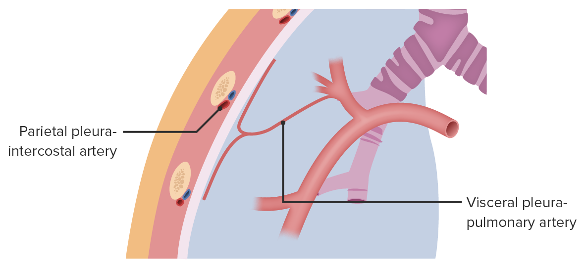
Blood supply of the pleurae:
The parietal pleura receives blood supply from the intercostal, diaphragmatic, mediastinal, and internal thoracic arteries. The visceral pleura receives blood supply from the bronchial and pulmonary vessels.

Image by Lecturio. License: CC BY-NC-SA 4.0

Clinical Relevance

Infectious disorders of the pleura

  • Pleural effusion Pleural Effusion Pleural effusion refers to the accumulation of fluid between the layers of the parietal and visceral pleura. Common causes of this condition include infection, malignancy, autoimmune disorders, or volume overload. Clinical manifestations include chest pain, cough, and dyspnea. Pleural Effusion: the accumulation of fluid between the layers of the parietal Parietal One of a pair of irregularly shaped quadrilateral bones situated between the frontal bone and occipital bone, which together form the sides of the cranium. Skull: Anatomy and visceral pleura. Caused by infection, malignancy Malignancy Hemothorax, autoimmune disorders, or volume overload. Presents as chest pain Pain An unpleasant sensation induced by noxious stimuli which are detected by nerve endings of nociceptive neurons. Pain: Types and Pathways, cough, and dyspnea Dyspnea Dyspnea is the subjective sensation of breathing discomfort. Dyspnea is a normal manifestation of heavy physical or psychological exertion, but also may be caused by underlying conditions (both pulmonary and extrapulmonary). Dyspnea. Classified as transudates or exudates, pleural effusions are usually diagnosed clinically, although imaging can confirm the diagnosis. Management is dependent on the underlying condition.
  • Pleuritis Pleuritis Pleuritis, also known as pleurisy, is an inflammation of the visceral and parietal layers of the pleural membranes of the lungs. The condition can be primary or secondary and results in sudden, sharp, and intense chest pain on inhalation and exhalation. Pleuritis: also known as pleurisy Pleurisy Pleuritis, also known as pleurisy, is an inflammation of the visceral and parietal layers of the pleural membranes of the lungs. The condition can be primary or secondary and results in sudden, sharp, and intense chest pain on inhalation and exhalation. Pleuritis, an inflammation Inflammation Inflammation is a complex set of responses to infection and injury involving leukocytes as the principal cellular mediators in the body’s defense against pathogenic organisms. Inflammation is also seen as a response to tissue injury in the process of wound healing. The 5 cardinal signs of inflammation are pain, heat, redness, swelling, and loss of function. Inflammation of the pleura. Results in sudden and intense chest pain Pain An unpleasant sensation induced by noxious stimuli which are detected by nerve endings of nociceptive neurons. Pain: Types and Pathways on inhalation and exhalation, and usually presents as part of pneumonia Pneumonia Pneumonia or pulmonary inflammation is an acute or chronic inflammation of lung tissue. Causes include infection with bacteria, viruses, or fungi. In more rare cases, pneumonia can also be caused through toxic triggers through inhalation of toxic substances, immunological processes, or in the course of radiotherapy. Pneumonia. The pain Pain An unpleasant sensation induced by noxious stimuli which are detected by nerve endings of nociceptive neurons. Pain: Types and Pathways intensifies upon deep inspiration Inspiration Ventilation: Mechanics of Breathing or coughing. Caused by infection, trauma, cardiac ischemia Ischemia A hypoperfusion of the blood through an organ or tissue caused by a pathologic constriction or obstruction of its blood vessels, or an absence of blood circulation. Ischemic Cell Damage, and lung cancer Lung cancer Lung cancer is the malignant transformation of lung tissue and the leading cause of cancer-related deaths. The majority of cases are associated with long-term smoking. The disease is generally classified histologically as either small cell lung cancer or non-small cell lung cancer. Symptoms include cough, dyspnea, weight loss, and chest discomfort. Lung Cancer. Management consists of pain Pain An unpleasant sensation induced by noxious stimuli which are detected by nerve endings of nociceptive neurons. Pain: Types and Pathways control and the treatment of the underlying condition.

Traumatic disorders of the pleura

  • Pneumothorax Pneumothorax A pneumothorax is a life-threatening condition in which air collects in the pleural space, causing partial or full collapse of the lung. A pneumothorax can be traumatic or spontaneous. Patients present with a sudden onset of sharp chest pain, dyspnea, and diminished breath sounds on exam. Pneumothorax: a life-threatening condition in which air collects in the pleural space Pleural space The thin serous membrane enveloping the lungs (lung) and lining the thoracic cavity. Pleura consist of two layers, the inner visceral pleura lying next to the pulmonary parenchyma and the outer parietal pleura. Between the two layers is the pleural cavity which contains a thin film of liquid. Pleuritis, causing a partial or full collapse of the lung. Can be traumatic or spontaneous. Patients Patients Individuals participating in the health care system for the purpose of receiving therapeutic, diagnostic, or preventive procedures. Clinician–Patient Relationship present with a sudden onset of sharp chest pain Sharp Chest Pain Chest Pain, dyspnea Dyspnea Dyspnea is the subjective sensation of breathing discomfort. Dyspnea is a normal manifestation of heavy physical or psychological exertion, but also may be caused by underlying conditions (both pulmonary and extrapulmonary). Dyspnea, and diminished breath sounds. Diagnosis is made with imaging, although tension pneumothorax Tension Pneumothorax Pneumothorax is a clinical diagnosis. Management is based on its size and the stability of the patient.
  • Hemothorax Hemothorax A hemothorax is a collection of blood in the pleural cavity. Hemothorax most commonly occurs due to damage to the intercostal arteries or from a lung laceration following chest trauma. Hemothorax can also occur as a complication of disease, or hemothorax may be spontaneous or iatrogenic. Hemothorax: a collection of blood in the pleural cavity. Most commonly due to damage to the intercostal arteries Arteries Arteries are tubular collections of cells that transport oxygenated blood and nutrients from the heart to the tissues of the body. The blood passes through the arteries in order of decreasing luminal diameter, starting in the largest artery (the aorta) and ending in the small arterioles. Arteries are classified into 3 types: large elastic arteries, medium muscular arteries, and small arteries and arterioles. Arteries: Histology from chest trauma. Presents with shortness of breath Shortness of breath Dyspnea is the subjective sensation of breathing discomfort. Dyspnea is a normal manifestation of heavy physical or psychological exertion, but also may be caused by underlying conditions (both pulmonary and extrapulmonary). Dyspnea and chest pain Pain An unpleasant sensation induced by noxious stimuli which are detected by nerve endings of nociceptive neurons. Pain: Types and Pathways. Physical exam findings include hypotension Hypotension Hypotension is defined as low blood pressure, specifically < 90/60 mm Hg, and is most commonly a physiologic response. Hypotension may be mild, serious, or life threatening, depending on the cause. Hypotension, tachycardia Tachycardia Abnormally rapid heartbeat, usually with a heart rate above 100 beats per minute for adults. Tachycardia accompanied by disturbance in the cardiac depolarization (cardiac arrhythmia) is called tachyarrhythmia. Sepsis in Children, decreased lung sounds, and dullness on percussion Percussion Act of striking a part with short, sharp blows as an aid in diagnosing the condition beneath the sound obtained. Pulmonary Examination of the chest. Diagnosis is by upright chest X-ray X-ray Penetrating electromagnetic radiation emitted when the inner orbital electrons of an atom are excited and release radiant energy. X-ray wavelengths range from 1 pm to 10 nm. Hard x-rays are the higher energy, shorter wavelength x-rays. Soft x-rays or grenz rays are less energetic and longer in wavelength. The short wavelength end of the x-ray spectrum overlaps the gamma rays wavelength range. The distinction between gamma rays and x-rays is based on their radiation source. Pulmonary Function Tests. Management is usually with tube thoracostomy Tube Thoracostomy Surgical procedure involving the creation of an opening (stoma) into the chest cavity for drainage; used in the treatment of pleural effusion; pneumothorax; hemothorax; and empyema. Thoracic Surgery drainage. Thoracoscopic surgery or thoracotomy Thoracotomy Surgical incision into the chest wall. Thoracic Surgery may be indicated in specific circumstances.

References

  1. Drake, R.L., Vogl, A.W., & Mitchell, A.W.M. (2014). Gray’s Anatomy for Students (3rd ed.). Philadelphia, PA:  Churchill Livingstone. 
  2. Standring, S. (2016). Gray’s anatomy. The Anatomical Basis of Clinical Practice (41st ed.). Edinburgh: Churchill Livingstone/Elsevier.

Create your free account or log in to continue reading!

Sign up now and get free access to Lecturio with concept pages, medical videos, and questions for your medical education.

User Reviews

Details