Achieve Mastery of Medical Concepts

Study for medical school and boards with Lecturio

Paranasal Sinuses: Anatomy

The 4 pair of paranasal sinuses include the maxillary, ethmoid, sphenoid, and frontal Frontal The bone that forms the frontal aspect of the skull. Its flat part forms the forehead, articulating inferiorly with the nasal bone and the cheek bone on each side of the face. Skull: Anatomy sinuses. The sinuses are a group of air-filled cavities located within the facial and cranial skeleton; all are connected to the main nasal cavity Nasal cavity The proximal portion of the respiratory passages on either side of the nasal septum. Nasal cavities, extending from the nares to the nasopharynx, are lined with ciliated nasal mucosa. Nose Anatomy (External & Internal) and nasopharynx Nasopharynx The top portion of the pharynx situated posterior to the nose and superior to the soft palate. The nasopharynx is the posterior extension of the nasal cavities and has a respiratory function. Pharynx: Anatomy. Functions include contributing to voice resonance, reducing the skull Skull The skull (cranium) is the skeletal structure of the head supporting the face and forming a protective cavity for the brain. The skull consists of 22 bones divided into the viscerocranium (facial skeleton) and the neurocranium. Skull: Anatomy weight to facilitate an upright head position, conditioning (warming and humidifying) inhaled air, and maximizing the surface of the nasal mucosa Nasal mucosa The mucous lining of the nasal cavity, including lining of the nostril (vestibule) and the olfactory mucosa. Nasal mucosa consists of ciliated cells, goblet cells, brush cells, small granule cells, basal cells (stem cells) and glands containing both mucous and serous cells. Nose Anatomy (External & Internal).

Last updated: Jan 11, 2024

Editorial responsibility: Stanley Oiseth, Lindsay Jones, Evelin Maza

Maxillary Sinuses

Overview

  • Contained in the maxillary bone Maxillary bone One of a pair of irregularly shaped bones that form the upper jaw. A maxillary bone provides tooth sockets for the superior teeth, forms part of the orbit, and contains the maxillary sinus. Skull: Anatomy below the eyes
  • The largest of the paranasal sinuses
  • Pyramid-shaped sinus
  • Drains into the middle nasal meatus Nasal meatus Nose Anatomy (External & Internal)
  • Present at birth and fully pneumatized (air-filled) by the age of 7

Relations

  • Medial wall: the lateral wall of the main nasal cavity Nasal cavity The proximal portion of the respiratory passages on either side of the nasal septum. Nasal cavities, extending from the nares to the nasopharynx, are lined with ciliated nasal mucosa. Nose Anatomy (External & Internal)
  • Roof: the floor of the orbit
  • Anterior wall: contains the infraorbital foramen and houses the infraorbital nerve
  • Anterolateral walls: lateral maxilla Maxilla One of a pair of irregularly shaped bones that form the upper jaw. A maxillary bone provides tooth sockets for the superior teeth, forms part of the orbit, and contains the maxillary sinus. Skull: Anatomy
  • Floor: the alveolar process of the maxilla Maxilla One of a pair of irregularly shaped bones that form the upper jaw. A maxillary bone provides tooth sockets for the superior teeth, forms part of the orbit, and contains the maxillary sinus. Skull: Anatomy (close to the roots of the 4 molars Molars The most posterior teeth on either side of the jaw, totaling eight in the deciduous dentition (2 on each side, upper and lower), and usually 12 in the permanent dentition (three on each side, upper and lower). They are grinding teeth, having large crowns and broad chewing surfaces. Teeth: Anatomy)
  • Posterior wall: borders the pterygopalatine and infratemporal fossa Infratemporal fossa The compartment that lies posterior to the maxilla and inferior to the side wall of the skull deep to the ramus of the mandible. It contains several facial nerves and the carotid arteries and the jugular veins. Skull: Anatomy

Neurovasculature

  • Lymphatic drainage: submandibular lymph nodes Lymph Nodes They are oval or bean shaped bodies (1 – 30 mm in diameter) located along the lymphatic system. Lymphatic Drainage System: Anatomy 
  • Blood supply (branches of the internal maxillary artery):
    • Anterior superior alveolar artery
    • Middle superior alveolar artery
    • Posterior superior alveolar artery
    • Infraorbital artery
    • Sphenopalatine artery
    • Greater palatine artery Greater palatine artery Palate: Anatomy (branch of the descending palatine artery)
  • Innervation:
    • Maxillary branches (V2) of the trigeminal nerve Trigeminal nerve The 5th and largest cranial nerve. The trigeminal nerve is a mixed motor and sensory nerve. The larger sensory part forms the ophthalmic, mandibular, and maxillary nerves which carry afferents sensitive to external or internal stimuli from the skin, muscles, and joints of the face and mouth and from the teeth. Most of these fibers originate from cells of the trigeminal ganglion and project to the trigeminal nucleus of the brain stem. The smaller motor part arises from the brain stem trigeminal motor nucleus and innervates the muscles of mastication. The 12 Cranial Nerves: Overview and Functions
    • Infraorbital nerve
    • Greater palatine nerve Greater palatine nerve Palate: Anatomy(s)
Sinusitis

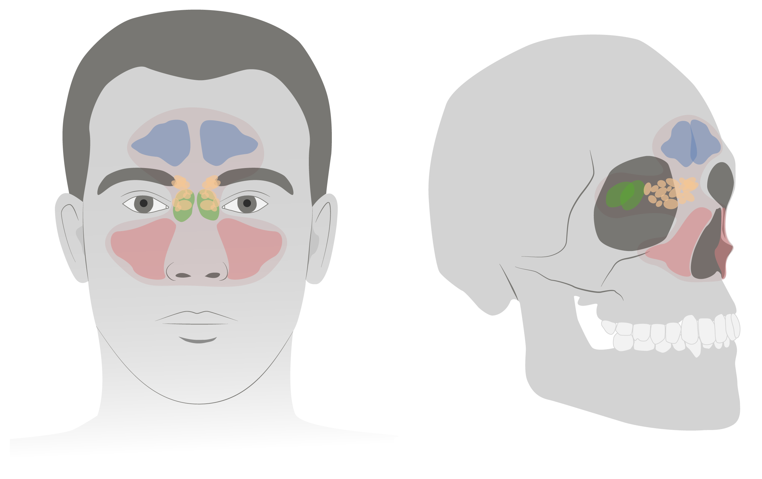
The maxillary sinuses (pink) are pyramid-shaped sinuses within the cheeks.

Image by Lecturio.

Frontal Sinuses

Overview

  • Contained in the frontal Frontal The bone that forms the frontal aspect of the skull. Its flat part forms the forehead, articulating inferiorly with the nasal bone and the cheek bone on each side of the face. Skull: Anatomy bone Bone Bone is a compact type of hardened connective tissue composed of bone cells, membranes, an extracellular mineralized matrix, and central bone marrow. The 2 primary types of bone are compact and spongy. Bones: Structure and Types of the forehead Forehead The part of the face above the eyes. Melasma above the eyes
  • Large, irregularly shaped, chambered sinus (can be congenitally hypoplastic/aplastic)
  • Appears at the age of 6 and fully develops during adulthood
  • Drains via the frontonasal duct into the semilunar hiatus in the middle nasal meatus Nasal meatus Nose Anatomy (External & Internal)

Relations

  • Floor:
    • Orbital roof (danger of perforation Perforation A pathological hole in an organ, blood vessel or other soft part of the body, occurring in the absence of external force. Esophagitis into the orbital cavity in sinusitis Sinusitis Sinusitis refers to inflammation of the mucosal lining of the paranasal sinuses. The condition usually occurs concurrently with inflammation of the nasal mucosa (rhinitis), a condition known as rhinosinusitis. Acute sinusitis is due to an upper respiratory infection caused by a viral, bacterial, or fungal agent. Sinusitis)
    • Anterior ethmoidal sinuses
    • Nasal cavity Nasal cavity The proximal portion of the respiratory passages on either side of the nasal septum. Nasal cavities, extending from the nares to the nasopharynx, are lined with ciliated nasal mucosa. Nose Anatomy (External & Internal)
  • Superoposterior wall: base of the skull Base of the skull The inferior region of the skull consisting of an internal (cerebral), and an external (basilar) surface. Skull: Anatomy (danger of intracranial infection) 
  • Anterior wall: superciliary arches

Neurovasculature

  • Lymphatic drainage: submandibular lymph nodes Lymph Nodes They are oval or bean shaped bodies (1 – 30 mm in diameter) located along the lymphatic system. Lymphatic Drainage System: Anatomy
  • Blood supply (branches of the internal carotid artery Internal carotid artery Branch of the common carotid artery which supplies the anterior part of the brain, the eye and its appendages, the forehead and nose. Carotid Arterial System: Anatomy via the ophthalmic artery Ophthalmic artery Artery originating from the internal carotid artery and distributing to the eye, orbit and adjacent facial structures. Eye: Anatomy):
    • Anterior ethmoidal artery
    • Supraorbital artery
    • Supratrochlear artery
  • Innervation (branches of the ophthalmic nerve (V1)):
    • Supraorbital nerve
    • Supratrochlear nerve
Sinusitis

The frontal sinuses (blue) are irregularly shaped sinuses above the orbital cavities.

Image by Lecturio.

Ethmoid Sinuses

Overview

Relations

  • Superior wall: floor of anterior cranial fossa Anterior cranial fossa The compartment containing the inferior part and anterior extremities of the frontal lobes (frontal lobe) of the cerebral hemispheres. It is formed mainly by orbital parts of the frontal bone and the lesser wings of the sphenoid bone. Skull: Anatomy and frontal Frontal The bone that forms the frontal aspect of the skull. Its flat part forms the forehead, articulating inferiorly with the nasal bone and the cheek bone on each side of the face. Skull: Anatomy bone Bone Bone is a compact type of hardened connective tissue composed of bone cells, membranes, an extracellular mineralized matrix, and central bone marrow. The 2 primary types of bone are compact and spongy. Bones: Structure and Types
  • Lateral walls: medial wall of the orbital cavity 
  • Medial walls: superolateral walls of the nasal cavity Nasal cavity The proximal portion of the respiratory passages on either side of the nasal septum. Nasal cavities, extending from the nares to the nasopharynx, are lined with ciliated nasal mucosa. Nose Anatomy (External & Internal) and the middle turbinate

Neurovasculature

  • Lymphatic drainage:
  • Blood supply:
    • Anterior and posterior ethmoidal arteries Arteries Arteries are tubular collections of cells that transport oxygenated blood and nutrients from the heart to the tissues of the body. The blood passes through the arteries in order of decreasing luminal diameter, starting in the largest artery (the aorta) and ending in the small arterioles. Arteries are classified into 3 types: large elastic arteries, medium muscular arteries, and small arteries and arterioles. Arteries: Histology (branches of ophthalmic artery Ophthalmic artery Artery originating from the internal carotid artery and distributing to the eye, orbit and adjacent facial structures. Eye: Anatomy)
    • Sphenopalatine artery (branch of external carotid artery External carotid artery Branch of the common carotid artery which supplies the exterior of the head, the face, and the greater part of the neck. Carotid Arterial System: Anatomy)
  • Innervation:
    • Anterior and posterior ethmoidal nerves
    • Posterior lateral superior and inferior nasal nerves
    • Orbital branches of the pterygopalatine ganglion
Sinusitis

The ethmoid sinuses (yellow) are thin-walled sinuses medial to the optic cavities.

Image by Lecturio.

Sphenoidal Sinuses

Overview

  • Contained in the sphenoid bone Sphenoid bone An irregular unpaired bone situated at the skull base and wedged between the frontal, temporal, and occipital bones (frontal bone; temporal bone; occipital bone). Sphenoid bone consists of a median body and three pairs of processes resembling a bat with spread wings. The body is hollowed out in its inferior to form two large cavities (sphenoid sinus). Orbit and Extraocular Muscles: Anatomy centrally located in the skull Skull The skull (cranium) is the skeletal structure of the head supporting the face and forming a protective cavity for the brain. The skull consists of 22 bones divided into the viscerocranium (facial skeleton) and the neurocranium. Skull: Anatomy
  • The most posterior sinus: variable Variable Variables represent information about something that can change. The design of the measurement scales, or of the methods for obtaining information, will determine the data gathered and the characteristics of that data. As a result, a variable can be qualitative or quantitative, and may be further classified into subgroups. Types of Variables shape and size
  • Rarely symmetrical Symmetrical Dermatologic Examination
  • Drains into the sphenoethmoidal recess medial to the superior turbinate
  • Appears at the age of 3 and fully develops during adulthood

Relations

  • Floor: roof of the nasopharynx Nasopharynx The top portion of the pharynx situated posterior to the nose and superior to the soft palate. The nasopharynx is the posterior extension of the nasal cavities and has a respiratory function. Pharynx: Anatomy and pterygoid canal 
  • Anterior wall: posterior wall of the nasal cavity Nasal cavity The proximal portion of the respiratory passages on either side of the nasal septum. Nasal cavities, extending from the nares to the nasopharynx, are lined with ciliated nasal mucosa. Nose Anatomy (External & Internal)
  • Roof: (close proximity to):
    • Sella turcica Sella turcica A bony prominence situated on the upper surface of the body of the sphenoid bone. It houses the pituitary gland. Pituitary Gland: Anatomy
    • Pituitary Pituitary A small, unpaired gland situated in the sella turcica. It is connected to the hypothalamus by a short stalk which is called the infundibulum. Hormones: Overview and Types gland
    • Optic chiasm Optic Chiasm The x-shaped structure formed by the meeting of the two optic nerves. At the optic chiasm the fibers from the medial part of each retina cross to project to the other side of the brain while the lateral retinal fibers continue on the same side. As a result each half of the brain receives information about the contralateral visual field from both eyes. The Visual Pathway and Related Disorders
    • Front and middle cranial fossa Middle cranial fossa The compartment containing the anterior extremities and half the inferior surface of the temporal lobes (temporal lobe) of the cerebral hemispheres. Lying posterior and inferior to the anterior cranial fossa, it is formed by part of the temporal bone and sphenoid bone. It is separated from the posterior cranial fossa by crests formed by the superior borders of the petrous parts of the temporal bones. Skull: Anatomy
  • Lateral walls: proximity to:
  • Medial wall: septum of the sphenoid bone Sphenoid bone An irregular unpaired bone situated at the skull base and wedged between the frontal, temporal, and occipital bones (frontal bone; temporal bone; occipital bone). Sphenoid bone consists of a median body and three pairs of processes resembling a bat with spread wings. The body is hollowed out in its inferior to form two large cavities (sphenoid sinus). Orbit and Extraocular Muscles: Anatomy

Neurovasculature

  • Lymphatic drainage: retropharyngeal lymph nodes Lymph Nodes They are oval or bean shaped bodies (1 – 30 mm in diameter) located along the lymphatic system. Lymphatic Drainage System: Anatomy
  • Blood supply:
    • Sphenopalatine artery (branch of external carotid artery External carotid artery Branch of the common carotid artery which supplies the exterior of the head, the face, and the greater part of the neck. Carotid Arterial System: Anatomy)
    • Posterior ethmoidal artery (branch of ophthalmic artery Ophthalmic artery Artery originating from the internal carotid artery and distributing to the eye, orbit and adjacent facial structures. Eye: Anatomy)
  • Innervation: 1st and 2nd divisions of the trigeminal nerve Trigeminal nerve The 5th and largest cranial nerve. The trigeminal nerve is a mixed motor and sensory nerve. The larger sensory part forms the ophthalmic, mandibular, and maxillary nerves which carry afferents sensitive to external or internal stimuli from the skin, muscles, and joints of the face and mouth and from the teeth. Most of these fibers originate from cells of the trigeminal ganglion and project to the trigeminal nucleus of the brain stem. The smaller motor part arises from the brain stem trigeminal motor nucleus and innervates the muscles of mastication. The 12 Cranial Nerves: Overview and Functions (V1 and V2)
Sinusitis

The sphenoidal sinuses (green) are located posterior to the ethmoid sinuses (yellow).

Image by Lecturio.

Clinical Relevance

  • Sinusitis Sinusitis Sinusitis refers to inflammation of the mucosal lining of the paranasal sinuses. The condition usually occurs concurrently with inflammation of the nasal mucosa (rhinitis), a condition known as rhinosinusitis. Acute sinusitis is due to an upper respiratory infection caused by a viral, bacterial, or fungal agent. Sinusitis: inflammation Inflammation Inflammation is a complex set of responses to infection and injury involving leukocytes as the principal cellular mediators in the body’s defense against pathogenic organisms. Inflammation is also seen as a response to tissue injury in the process of wound healing. The 5 cardinal signs of inflammation are pain, heat, redness, swelling, and loss of function. Inflammation of the mucous membrane Mucous membrane An epithelium with mucus-secreting cells, such as goblet cells. It forms the lining of many body cavities, such as the digestive tract, the respiratory tract, and the reproductive tract. Mucosa, rich in blood and lymph vessels, comprises an inner epithelium, a middle layer (lamina propria) of loose connective tissue, and an outer layer (muscularis mucosae) of smooth muscle cells that separates the mucosa from submucosa. Barrett Esophagus of the paranasal sinuses. Sinusitis Sinusitis Sinusitis refers to inflammation of the mucosal lining of the paranasal sinuses. The condition usually occurs concurrently with inflammation of the nasal mucosa (rhinitis), a condition known as rhinosinusitis. Acute sinusitis is due to an upper respiratory infection caused by a viral, bacterial, or fungal agent. Sinusitis is extremely common and usually presents concurrently with rhinitis Rhinitis Inflammation of the nasal mucosa, the mucous membrane lining the nasal cavities. Rhinitis ( inflammation Inflammation Inflammation is a complex set of responses to infection and injury involving leukocytes as the principal cellular mediators in the body’s defense against pathogenic organisms. Inflammation is also seen as a response to tissue injury in the process of wound healing. The 5 cardinal signs of inflammation are pain, heat, redness, swelling, and loss of function. Inflammation of the nasal mucosa Nasal mucosa The mucous lining of the nasal cavity, including lining of the nostril (vestibule) and the olfactory mucosa. Nasal mucosa consists of ciliated cells, goblet cells, brush cells, small granule cells, basal cells (stem cells) and glands containing both mucous and serous cells. Nose Anatomy (External & Internal)). The cause can be a viral, bacterial, or fungal infection, or an allergic reaction. Sinusitis Sinusitis Sinusitis refers to inflammation of the mucosal lining of the paranasal sinuses. The condition usually occurs concurrently with inflammation of the nasal mucosa (rhinitis), a condition known as rhinosinusitis. Acute sinusitis is due to an upper respiratory infection caused by a viral, bacterial, or fungal agent. Sinusitis can present as either acute or chronic inflammation Chronic Inflammation Inflammation and most commonly affects the maxillary sinuses. 
  • Choanal atresia Atresia Hypoplastic Left Heart Syndrome (HLHS): a congenital Congenital Chorioretinitis condition characterized by an obstruction of the posterior nasal aperture leading into the nasopharynx Nasopharynx The top portion of the pharynx situated posterior to the nose and superior to the soft palate. The nasopharynx is the posterior extension of the nasal cavities and has a respiratory function. Pharynx: Anatomy, which can manifest unilaterally or bilaterally. Bilateral choanal atresia Atresia Hypoplastic Left Heart Syndrome (HLHS) manifests as obstructed nasal breathing with intermittent cyanosis Cyanosis A bluish or purplish discoloration of the skin and mucous membranes due to an increase in the amount of deoxygenated hemoglobin in the blood or a structural defect in the hemoglobin molecule. Pulmonary Examination immediately after birth.
  • Mucormycosis Mucormycosis Mucormycosis is an angioinvasive fungal infection caused by multiple fungi within the order, Mucorales. The fungi are ubiquitous in the environment, but mucormycosis is very rare and almost always occurs in patients who are immunocompromised. Inhalation of fungal spores can cause rhinocerebral or pulmonary mucormycosis, direct inoculation can cause cutaneous mucormycosis, and ingestion can cause gastrointestinal mucormycosis. Mucorales/Mucormycosis: an angioinvasive fungal infection. Inhalation of fungal spores Spores The reproductive elements of lower organisms, such as bacteria; fungi; and cryptogamic plants. Anthrax can cause rhinocerebral mucormycosis Rhinocerebral mucormycosis Mucorales/Mucormycosis or pulmonary mucormycosis Pulmonary mucormycosis Mucorales/Mucormycosis. The clinical presentation results from fungal hyphae Hyphae Microscopic threadlike filaments in fungi that are filled with a layer of protoplasm. Collectively, the hyphae make up the mycelium. Mycology invading the blood vessels, causing thrombosis Thrombosis Formation and development of a thrombus or blood clot in the blood vessel. Epidemic Typhus and (ultimately) tissue necrosis Necrosis The death of cells in an organ or tissue due to disease, injury or failure of the blood supply. Ischemic Cell Damage. Diagnosis is confirmed with the identification Identification Defense Mechanisms of the organism on histopathology from biopsy Biopsy Removal and pathologic examination of specimens from the living body. Ewing Sarcoma specimens. Patients Patients Individuals participating in the health care system for the purpose of receiving therapeutic, diagnostic, or preventive procedures. Clinician–Patient Relationship must be treated aggressively with surgical resection of infected tissues and systemic antifungals. 

References

  1. Petrikkos, G., et al. (2012). Epidemiology and clinical manifestations of mucormycosis. Clin Infect Dis. 54 (Suppl 1), S23–34. https://www.doi.org/10.1093/cid/cir866
  2. Cox, G.M. (2021). Mucormycosis (zygomycosis). In Bond, S. (Ed.), UpToDate. Retrieved June 4, 2021, from https://www.uptodate.com/contents/mucormycosis-zygomycosis
  3. Isaacson, G.C. (2021). Congenital anomalies of the nose. In Messner, A. (Ed.), UpToDate. Retrieved July 2021, from https://www.uptodate.com/contents/congenital-anomalies-of-the-nose

Create your free account or log in to continue reading!

Sign up now and get free access to Lecturio with concept pages, medical videos, and questions for your medical education.

User Reviews

Details