The palate is the structure that forms the roof of the mouth and floor of the nasal cavity Nasal cavity The proximal portion of the respiratory passages on either side of the nasal septum. Nasal cavities, extending from the nares to the nasopharynx, are lined with ciliated nasal mucosa. Nose Anatomy (External & Internal). This structure is divided into soft and hard palates. The palate is formed between weeks 7 and 10 of gestation, and deformities of this structure ( cleft palate Cleft palate Congenital fissure of the soft and/or hard palate, due to faulty fusion. Cleft Lip and Cleft Palate) are usually relevant because of its role in feeding, especially in infants.
Last updated: Dec 15, 2025
The embryology of the palate is complex, with a critical stepwise process to midline fusion. Interruption of this process can result in cleft disorders.
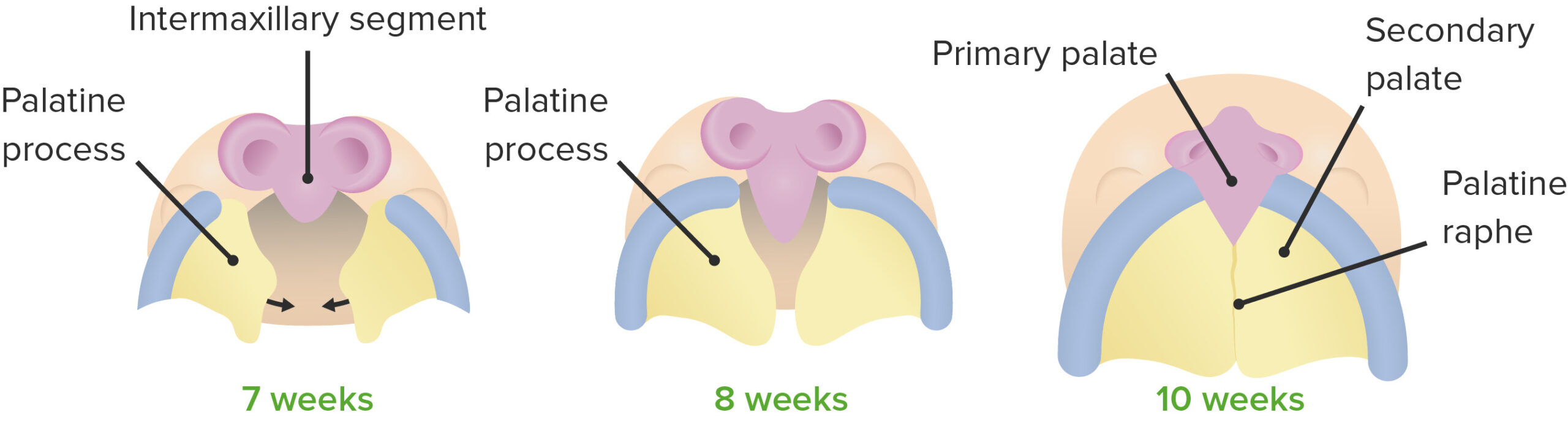
Formation of the palate
Image by Lecturio.The palate is divided into the hard palate and soft palate. The hard palate is located anterior to the soft palate, and each receives blood supply from branches of the external carotid artery External carotid artery Branch of the common carotid artery which supplies the exterior of the head, the face, and the greater part of the neck. Carotid Arterial System: Anatomy and nerve supply via the palatine and nasopalatine nerves.
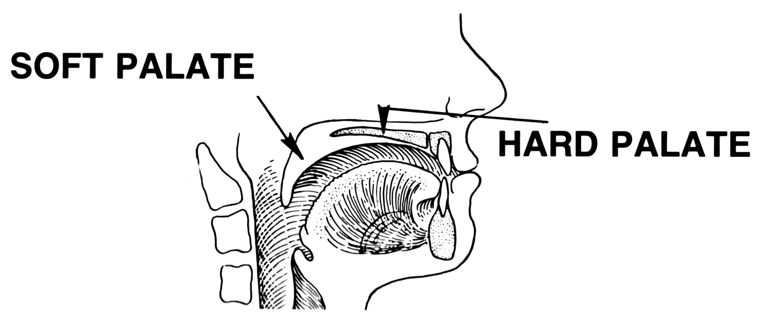
Location and relations between the hard and soft palate
Image: “Line art drawing of palate” by Pearson Scott Foresman. License: Public DomainHard palate (anterior):
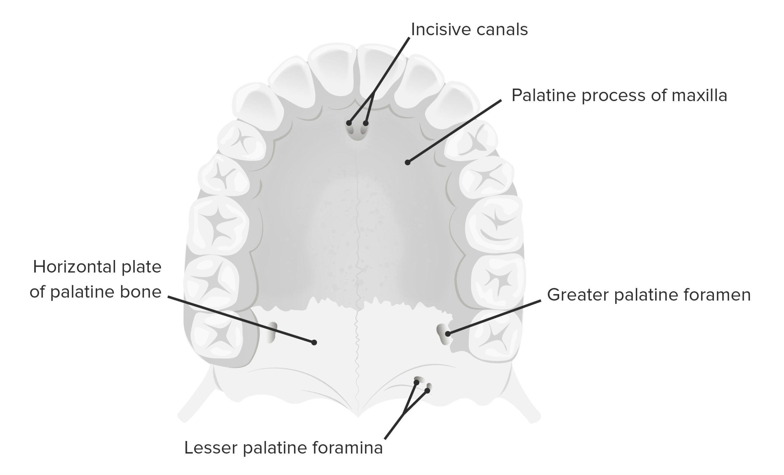
Inferior view of the hard palate, showcasing the 2 palatine processes of the maxilla and the horizontal plate of the palatine bones
Image by Lecturio.Soft palate (posterior):
| Muscle | Origin | Insertion | Nerve supply | Function |
|---|---|---|---|---|
| Musculus uvulae | Posterior nasal spine Spine The human spine, or vertebral column, is the most important anatomical and functional axis of the human body. It consists of 7 cervical vertebrae, 12 thoracic vertebrae, and 5 lumbar vertebrae and is limited cranially by the skull and caudally by the sacrum. Vertebral Column: Anatomy | Musculus uvulae of the opposite side | Pharyngeal branch of vagus nerve Vagus nerve The 10th cranial nerve. The vagus is a mixed nerve which contains somatic afferents (from skin in back of the ear and the external auditory meatus), visceral afferents (from the pharynx, larynx, thorax, and abdomen), parasympathetic efferents (to the thorax and abdomen), and efferents to striated muscle (of the larynx and pharynx). Pharynx: Anatomy | Pulls uvula Uvula A fleshy extension at the back of the soft palate that hangs above the opening of the throat. Peritonsillar Abscess forward |
| Tensor veli palatini | Cartilaginous part of the auditory tube | Palatine aponeurosis | Muscular branch of the mandibular nerve Mandibular nerve A branch of the trigeminal (5th cranial) nerve. The mandibular nerve carries motor fibers to the muscles of mastication and sensory fibers to the teeth and gingivae, the face in the region of the mandible, and parts of the dura. Jaw and Temporomandibular Joint: Anatomy | Tenses the soft palate |
| Levator veli palatini | Cartilaginous portion of the eustachian tube Eustachian tube A narrow passageway that connects the upper part of the throat to the tympanic cavity. Ear: Anatomy | Palatine aponeurosis | Pharyngeal branch of vagus nerve Vagus nerve The 10th cranial nerve. The vagus is a mixed nerve which contains somatic afferents (from skin in back of the ear and the external auditory meatus), visceral afferents (from the pharynx, larynx, thorax, and abdomen), parasympathetic efferents (to the thorax and abdomen), and efferents to striated muscle (of the larynx and pharynx). Pharynx: Anatomy | Elevates soft palate |
| Palatopharyngeus | Palatine aponeurosis and hard palate | Upper border of the thyroid Thyroid The thyroid gland is one of the largest endocrine glands in the human body. The thyroid gland is a highly vascular, brownish-red gland located in the visceral compartment of the anterior region of the neck. Thyroid Gland: Anatomy cartilage Cartilage Cartilage is a type of connective tissue derived from embryonic mesenchyme that is responsible for structural support, resilience, and the smoothness of physical actions. Perichondrium (connective tissue membrane surrounding cartilage) compensates for the absence of vasculature in cartilage by providing nutrition and support. Cartilage: Histology | Elevates larynx Larynx The larynx, also commonly called the voice box, is a cylindrical space located in the neck at the level of the C3-C6 vertebrae. The major structures forming the framework of the larynx are the thyroid cartilage, cricoid cartilage, and epiglottis. The larynx serves to produce sound (phonation), conducts air to the trachea, and prevents large molecules from reaching the lungs. Larynx: Anatomy and the pharynx Pharynx The pharynx is a component of the digestive system that lies posterior to the nasal cavity, oral cavity, and larynx. The pharynx can be divided into the oropharynx, nasopharynx, and laryngopharynx. Pharyngeal muscles play an integral role in vital processes such as breathing, swallowing, and speaking. Pharynx: Anatomy | |
| Palatoglossus | Palatine aponeurosis | Lateral aspect of the tongue Tongue The tongue, on the other hand, is a complex muscular structure that permits tasting and facilitates the process of mastication and communication. The blood supply of the tongue originates from the external carotid artery, and the innervation is through cranial nerves. Lips and Tongue: Anatomy | Elevates posterior part of the tongue Tongue The tongue, on the other hand, is a complex muscular structure that permits tasting and facilitates the process of mastication and communication. The blood supply of the tongue originates from the external carotid artery, and the innervation is through cranial nerves. Lips and Tongue: Anatomy |
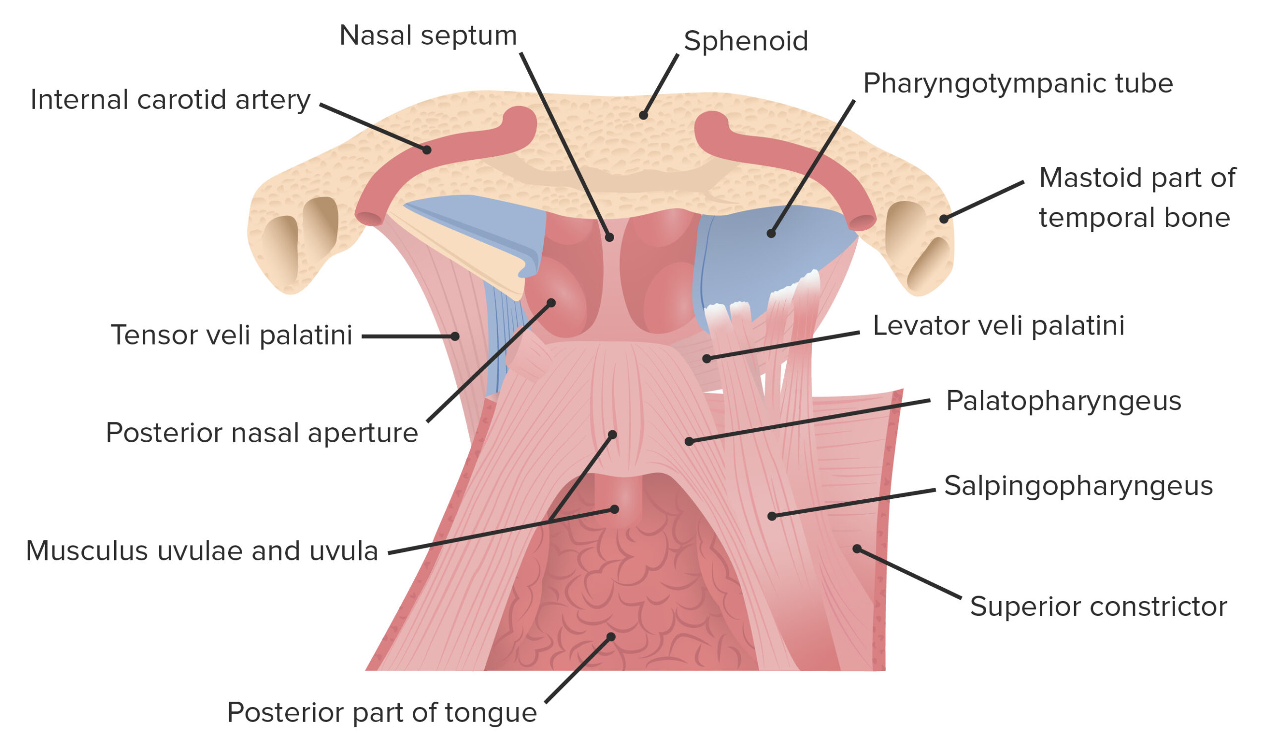
Muscles that comprise the soft palate:
This image is of the coronal plane viewed in an anteroposterior orientation.
Blood supply:
Innervation:
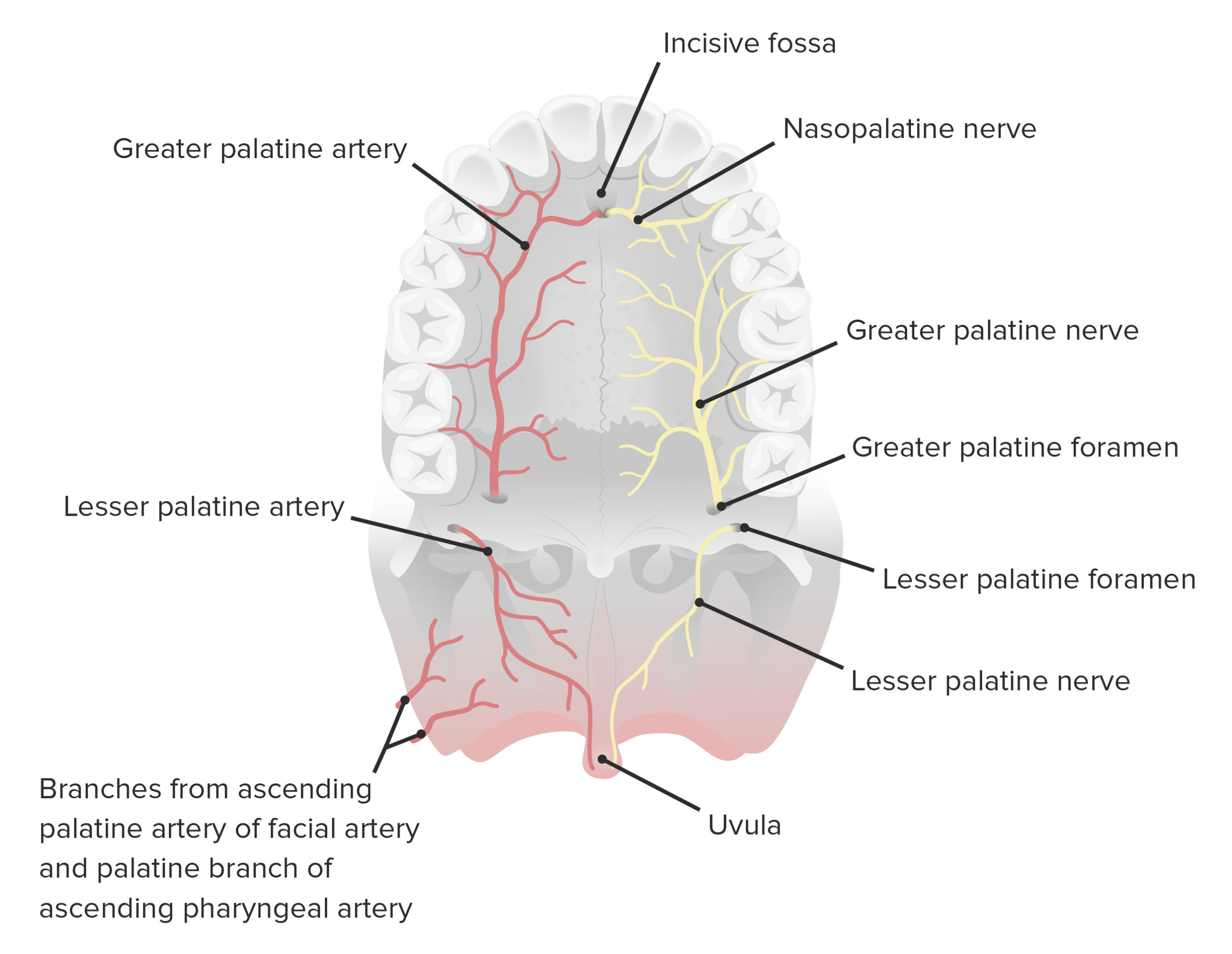
Blood supply and innervation of the palate
Image by Lecturio.The microscopic anatomy of the palate consists primarily of squamous epithelial tissue, although the hard palate also contains osseous components.
The palate contains:
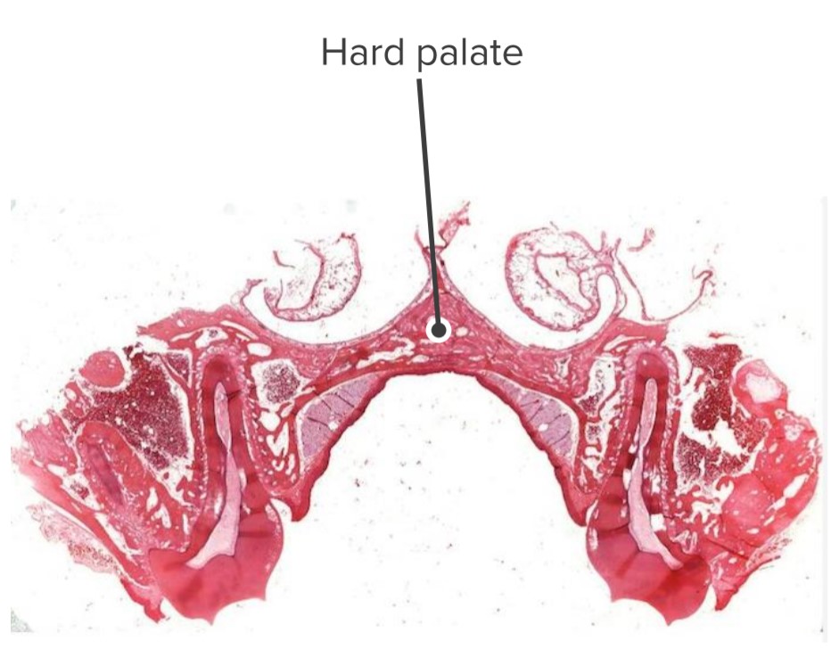
Hard palate and its relations with the nasal and oral cavities
Image by Geoffrey Meyer, PhD.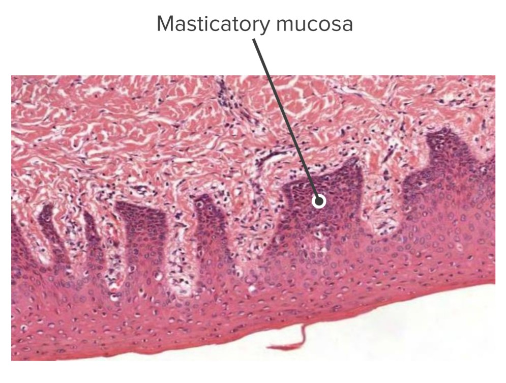
Masticatory mucosa of the palate
Image by Geoffrey Meyer, PhD.