Leukoplakia

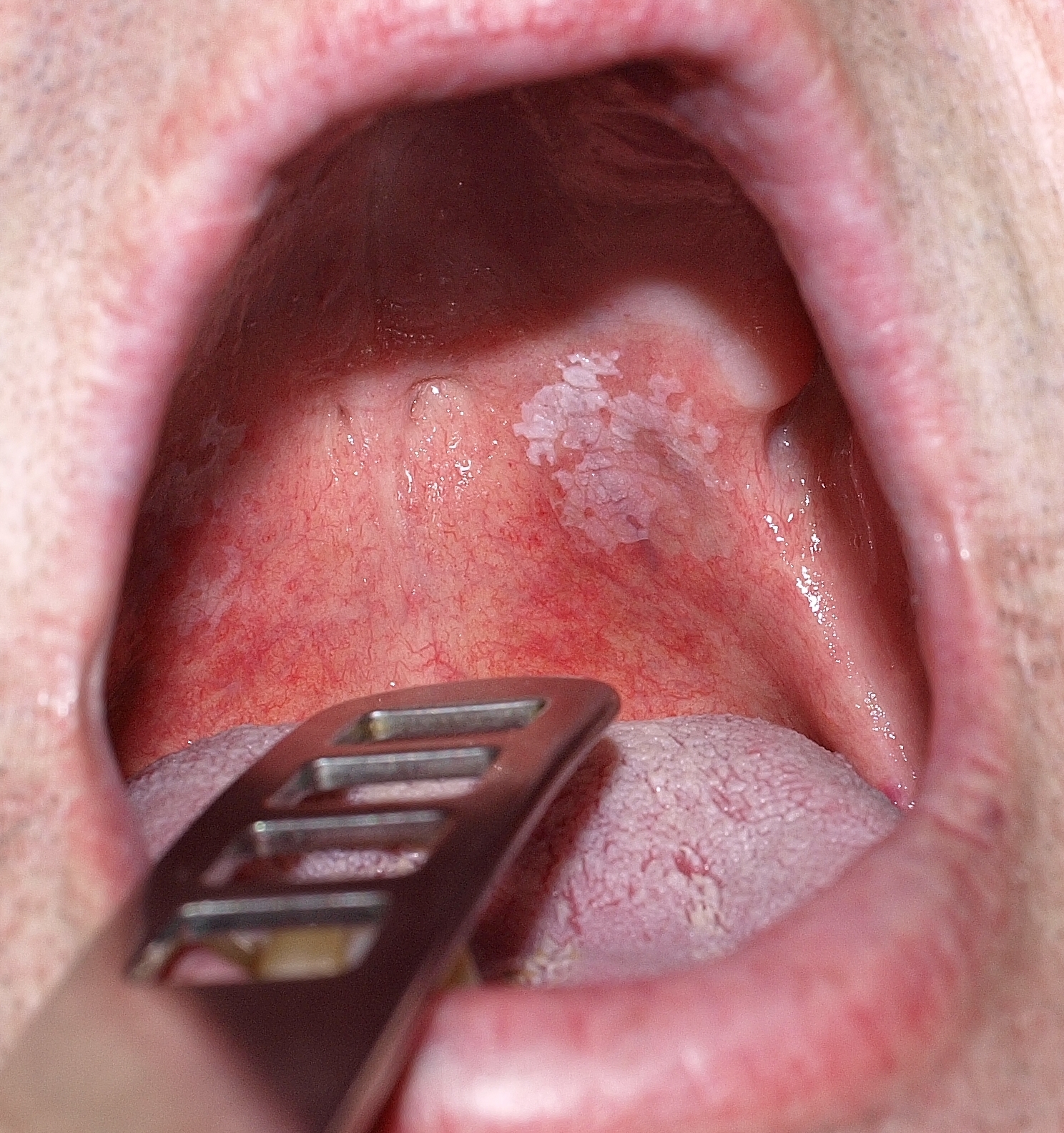
Leukoplakia is a potentially malignant lesion affecting the squamous epithelium Epithelium The epithelium is a complex of specialized cellular organizations arranged into sheets and lining cavities and covering the surfaces of the body. The cells exhibit polarity, having an apical and a basal pole. Structures important for the epithelial integrity and function involve the basement membrane, the semipermeable sheet on which the cells rest, and interdigitations, as well as cellular junctions. Surface Epithelium: Histology usually within the oral cavity. Leukoplakia can be associated with a history of chronic tobacco and alcohol use, both of which can synergistically damage the epithelium Epithelium The epithelium is a complex of specialized cellular organizations arranged into sheets and lining cavities and covering the surfaces of the body. The cells exhibit polarity, having an apical and a basal pole. Structures important for the epithelial integrity and function involve the basement membrane, the semipermeable sheet on which the cells rest, and interdigitations, as well as cellular junctions. Surface Epithelium: Histology. Leukoplakia presents as a white plaque Plaque Primary Skin Lesions that cannot be scraped off. Diagnosis is confirmed with a biopsy Biopsy Removal and pathologic examination of specimens from the living body. Ewing Sarcoma. The lesion can be surgically treated, but close observation is always recommended owing to the risk of malignant transformation Transformation Change brought about to an organism's genetic composition by unidirectional transfer (transfection; transduction, genetic; conjugation, genetic, etc.) and incorporation of foreign DNA into prokaryotic or eukaryotic cells by recombination of part or all of that DNA into the cell's genome. Bacteriology.

Last updated: Jul 1, 2026

Editorial responsibility: Stanley Oiseth, Lindsay Jones, Evelin Maza

Overview

Definition

Epidemiology

  • Prevalence Prevalence The total number of cases of a given disease in a specified population at a designated time. It is differentiated from incidence, which refers to the number of new cases in the population at a given time. Measures of Disease Frequency:
  • 80% of patients Patients Individuals participating in the health care system for the purpose of receiving therapeutic, diagnostic, or preventive procedures. Clinician–Patient Relationship are > 40 years of age.
  • Affects more men than women
  • Annual malignant transformation Transformation Change brought about to an organism’s genetic composition by unidirectional transfer (transfection; transduction, genetic; conjugation, genetic, etc.) and incorporation of foreign DNA into prokaryotic or eukaryotic cells by recombination of part or all of that DNA into the cell’s genome. Bacteriology rate: 1%–5%

Risk factors

Risk factors are similar to those for squamous cell carcinoma Squamous cell carcinoma Cutaneous squamous cell carcinoma (cSCC) is caused by malignant proliferation of atypical keratinocytes. This condition is the 2nd most common skin malignancy and usually affects sun-exposed areas of fair-skinned patients. The cancer presents as a firm, erythematous, keratotic plaque or papule. Squamous Cell Carcinoma (SCC).

  • Tobacco use (most common risk factor)
  • Alcohol consumption 
  • Increasing age
  • Infections Infections Invasion of the host organism by microorganisms or their toxins or by parasites that can cause pathological conditions or diseases. Chronic Granulomatous Disease:
    • HPV HPV Human papillomavirus (HPV) is a nonenveloped, circular, double-stranded DNA virus belonging to the Papillomaviridae family. Humans are the only reservoir, and transmission occurs through close skin-to-skin or sexual contact. Human papillomaviruses infect basal epithelial cells and can affect cell-regulatory proteins to result in cell proliferation. Papillomavirus (HPV)
    • Chronic candidiasis Candidiasis Candida is a genus of dimorphic, opportunistic fungi. Candida albicans is part of the normal human flora and is the most common cause of candidiasis. The clinical presentation varies and can include localized mucocutaneous infections (e.g., oropharyngeal, esophageal, intertriginous, and vulvovaginal candidiasis) and invasive disease (e.g., candidemia, intraabdominal abscess, pericarditis, and meningitis). Candida/Candidiasis

Pathophysiology

  • Exposure to carcinogens Carcinogens Substances that increase the risk of neoplasms in humans or animals. Both genotoxic chemicals, which affect DNA directly, and nongenotoxic chemicals, which induce neoplasms by other mechanism, are included. Carcinogenesis → genetic damage or mutations regulating cell turnover and death
  • Occurrence of cell proliferation and hyperkeratosis Hyperkeratosis Ichthyosis Vulgaris
  • Recurrent exposure to carcinogens Carcinogens Substances that increase the risk of neoplasms in humans or animals. Both genotoxic chemicals, which affect DNA directly, and nongenotoxic chemicals, which induce neoplasms by other mechanism, are included. Carcinogenesis → dysplasia

Clinical Presentation

General findings

  • Patches Patches Vitiligo or plaques:
    • Affect mucosa
    • Tend to be bright white
    • Cannot be scraped off 
  • Lesions are asymptomatic.

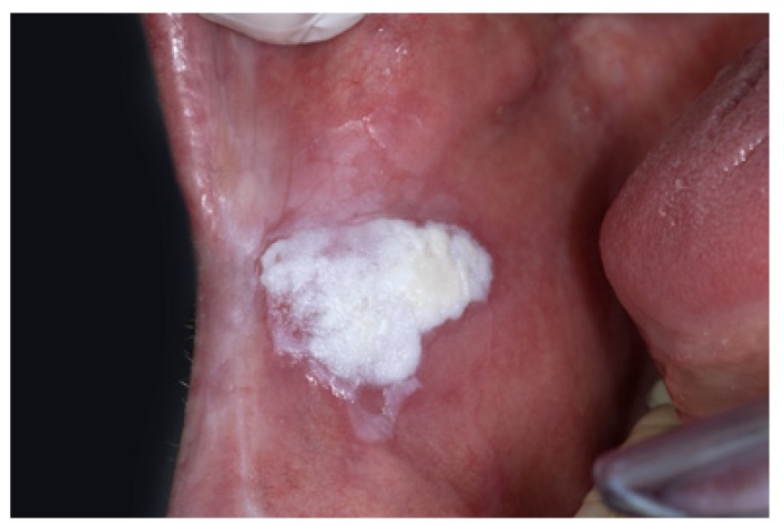
Homogeneous Homogeneous Imaging of the Spleen leukoplakia

This form is less likely to be malignant and is characterized by:
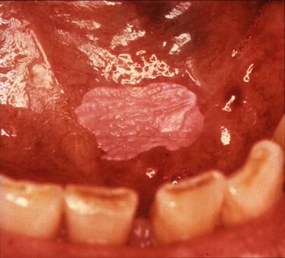
  • Uniformly white plaques 
  • Well-defined margins

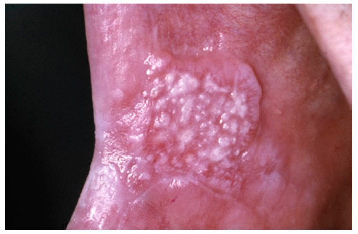
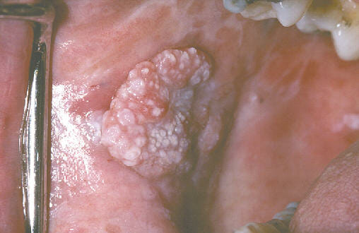
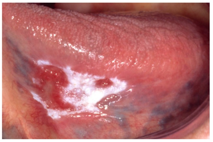
Nonhomogeneous leukoplakia

Nonhomogenous leukoplakia presents a higher risk of malignant transformation Transformation Change brought about to an organism’s genetic composition by unidirectional transfer (transfection; transduction, genetic; conjugation, genetic, etc.) and incorporation of foreign DNA into prokaryotic or eukaryotic cells by recombination of part or all of that DNA into the cell’s genome. Bacteriology and may appear:
Locations

  • Oral cavity (most common):
    • Tongue Tongue The tongue, on the other hand, is a complex muscular structure that permits tasting and facilitates the process of mastication and communication. The blood supply of the tongue originates from the external carotid artery, and the innervation is through cranial nerves. Lips and Tongue: Anatomy
    • Buccal mucosa Buccal mucosa Oral Cancer
  • Genitalia 
  • Esophagus Esophagus The esophagus is a muscular tube-shaped organ of around 25 centimeters in length that connects the pharynx to the stomach. The organ extends from approximately the 6th cervical vertebra to the 11th thoracic vertebra and can be divided grossly into 3 parts: the cervical part, the thoracic part, and the abdominal part. Esophagus: Anatomy 
  • Larynx Larynx The larynx, also commonly called the voice box, is a cylindrical space located in the neck at the level of the C3-C6 vertebrae. The major structures forming the framework of the larynx are the thyroid cartilage, cricoid cartilage, and epiglottis. The larynx serves to produce sound (phonation), conducts air to the trachea, and prevents large molecules from reaching the lungs. Larynx: Anatomy
  • Bladder Bladder A musculomembranous sac along the urinary tract. Urine flows from the kidneys into the bladder via the ureters, and is held there until urination. Pyelonephritis and Perinephric Abscess

Diagnosis and Management

Diagnosis

Biopsy Biopsy Removal and pathologic examination of specimens from the living body. Ewing Sarcoma is required for a definitive diagnosis, to evaluate for dysplasia, and to rule out other conditions. Findings include:
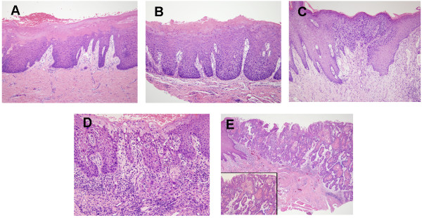
Histologic images of oral leukoplakia

Histological images of oral leukoplakia demonstrating progressive dysplasia and malignant transformation:
A: Hyperkeratosis with low-grade dysplasia present
B: Moderate dysplasia
C: High-grade dysplasia
D: Leukoplakia in the state of becoming invasive carcinoma
E: Invasive carcinoma

Image: “Representative histological pictures of OPLs and OSCCs” by Yang Yi et al. License: CC BY 2.0

Management

  • Close surveillance Surveillance Developmental Milestones and Normal Growth is indicated.
  • Elimination Elimination The initial damage and destruction of tumor cells by innate and adaptive immunity. Completion of the phase means no cancer growth. Cancer Immunotherapy of risk factors for all patients Patients Individuals participating in the health care system for the purpose of receiving therapeutic, diagnostic, or preventive procedures. Clinician–Patient Relationship, including smoking Smoking Willful or deliberate act of inhaling and exhaling smoke from burning substances or agents held by hand. Interstitial Lung Diseases and alcohol use
  • Medical therapy:
    • No routine medical therapy is recommended for oral leukoplakia Oral leukoplakia A white patch seen on the oral mucosa. It is considered a premalignant condition and is often tobacco-induced. When evidence of Epstein-Barr virus is present, the condition is called hairy leukoplakia. Oral Cancer.
    • In selected cases, specialists may consider topical therapy such as fluorouracil Fluorouracil A pyrimidine analog that is an antineoplastic antimetabolite. It interferes with DNA synthesis by blocking the thymidylate synthetase conversion of deoxyuridylic acid to thymidylic acid. Bowen Disease and Erythroplasia of Queyrat or imiquimod Imiquimod A topically-applied aminoquinoline immune modulator that induces interferon production. It is used in the treatment of external genital and perianal warts, superficial carcinoma, basal cell; and actinic keratosis. Hypertrophic and Keloid Scars.
  • Surgical management:
    • Options:
      • Surgical excision 
      • Laser ablation
      • Cryosurgery
    • Recommended for:
      • Lesions with moderate-to-severe dysplasia on biopsy Biopsy Removal and pathologic examination of specimens from the living body. Ewing Sarcoma 
      • Nonhomogeneous leukoplakia

Differential Diagnosis

  • Candidiasis Candidiasis Candida is a genus of dimorphic, opportunistic fungi. Candida albicans is part of the normal human flora and is the most common cause of candidiasis. The clinical presentation varies and can include localized mucocutaneous infections (e.g., oropharyngeal, esophageal, intertriginous, and vulvovaginal candidiasis) and invasive disease (e.g., candidemia, intraabdominal abscess, pericarditis, and meningitis). Candida/Candidiasis: a superficial fungal infection caused by Candida Candida Candida is a genus of dimorphic, opportunistic fungi. Candida albicans is part of the normal human flora and is the most common cause of candidiasis. The clinical presentation varies and can include localized mucocutaneous infections (e.g., oropharyngeal, esophageal, intertriginous, and vulvovaginal candidiasis) and invasive disease (e.g., candidemia, intraabdominal abscess, pericarditis, and meningitis). Candida/Candidiasis species. Candidiasis Candidiasis Candida is a genus of dimorphic, opportunistic fungi. Candida albicans is part of the normal human flora and is the most common cause of candidiasis. The clinical presentation varies and can include localized mucocutaneous infections (e.g., oropharyngeal, esophageal, intertriginous, and vulvovaginal candidiasis) and invasive disease (e.g., candidemia, intraabdominal abscess, pericarditis, and meningitis). Candida/Candidiasis can commonly occur on the oral mucosa Oral mucosa Lining of the oral cavity, including mucosa on the gums; the palate; the lip; the cheek; floor of the mouth; and other structures. The mucosa is generally a nonkeratinized stratified squamous epithelium covering muscle, bone, or glands but can show varying degree of keratinization at specific locations. Stomatitis, genitals, and skin Skin The skin, also referred to as the integumentary system, is the largest organ of the body. The skin is primarily composed of the epidermis (outer layer) and dermis (deep layer). The epidermis is primarily composed of keratinocytes that undergo rapid turnover, while the dermis contains dense layers of connective tissue. Skin: Structure and Functions folds. Infection of the oral mucosa Oral mucosa Lining of the oral cavity, including mucosa on the gums; the palate; the lip; the cheek; floor of the mouth; and other structures. The mucosa is generally a nonkeratinized stratified squamous epithelium covering muscle, bone, or glands but can show varying degree of keratinization at specific locations. Stomatitis presents as white plaques that bleed when scraped. The diagnosis is based on a clinical exam and can be confirmed by the identification Identification Defense Mechanisms of yeast Yeast A general term for single-celled rounded fungi that reproduce by budding. Brewers’ and bakers’ yeasts are saccharomyces cerevisiae; therapeutic dried yeast is yeast, dried. Mycology on a KOH wet mount. Candidiasis Candidiasis Candida is a genus of dimorphic, opportunistic fungi. Candida albicans is part of the normal human flora and is the most common cause of candidiasis. The clinical presentation varies and can include localized mucocutaneous infections (e.g., oropharyngeal, esophageal, intertriginous, and vulvovaginal candidiasis) and invasive disease (e.g., candidemia, intraabdominal abscess, pericarditis, and meningitis). Candida/Candidiasis can be treated with oral antifungal Antifungal Azoles medications.
  • Squamous cell carcinoma Squamous cell carcinoma Cutaneous squamous cell carcinoma (cSCC) is caused by malignant proliferation of atypical keratinocytes. This condition is the 2nd most common skin malignancy and usually affects sun-exposed areas of fair-skinned patients. The cancer presents as a firm, erythematous, keratotic plaque or papule. Squamous Cell Carcinoma (SCC): a condition caused by the malignant proliferation of atypical keratinocytes Keratinocytes Epidermal cells which synthesize keratin and undergo characteristic changes as they move upward from the basal layers of the epidermis to the cornified (horny) layer of the skin. Successive stages of differentiation of the keratinocytes forming the epidermal layers are basal cell, spinous or prickle cell, and the granular cell. Skin: Structure and Functions. Squamous cell carcinoma Squamous cell carcinoma Cutaneous squamous cell carcinoma (cSCC) is caused by malignant proliferation of atypical keratinocytes. This condition is the 2nd most common skin malignancy and usually affects sun-exposed areas of fair-skinned patients. The cancer presents as a firm, erythematous, keratotic plaque or papule. Squamous Cell Carcinoma (SCC) is the 2nd most common skin Skin The skin, also referred to as the integumentary system, is the largest organ of the body. The skin is primarily composed of the epidermis (outer layer) and dermis (deep layer). The epidermis is primarily composed of keratinocytes that undergo rapid turnover, while the dermis contains dense layers of connective tissue. Skin: Structure and Functions malignancy Malignancy Hemothorax and usually affects the sun-exposed areas of light-skinned patients Patients Individuals participating in the health care system for the purpose of receiving therapeutic, diagnostic, or preventive procedures. Clinician–Patient Relationship. Squamous cell carcinoma Squamous cell carcinoma Cutaneous squamous cell carcinoma (cSCC) is caused by malignant proliferation of atypical keratinocytes. This condition is the 2nd most common skin malignancy and usually affects sun-exposed areas of fair-skinned patients. The cancer presents as a firm, erythematous, keratotic plaque or papule. Squamous Cell Carcinoma (SCC) presents as a firm, erythematous, keratotic plaque Plaque Primary Skin Lesions or papule Papule Elevated lesion < 1 cm in diameter Generalized and Localized Rashes. A biopsy Biopsy Removal and pathologic examination of specimens from the living body. Ewing Sarcoma is used to confirm the diagnosis. Surgical excision is the mainstay of management.
  • Erythroplakia Erythroplakia A condition of the mucous membrane characterized by erythematous papular lesions. Oral Cancer: a precancerous Precancerous Pathological conditions that tend eventually to become malignant. Barrett Esophagus lesion that is similar to leukoplakia, which develops from chronic tobacco and alcohol use. Patients Patients Individuals participating in the health care system for the purpose of receiving therapeutic, diagnostic, or preventive procedures. Clinician–Patient Relationship usually present with granular, red, sharply demarcated lesions. A biopsy Biopsy Removal and pathologic examination of specimens from the living body. Ewing Sarcoma can show dysplastic keratinocytes Keratinocytes Epidermal cells which synthesize keratin and undergo characteristic changes as they move upward from the basal layers of the epidermis to the cornified (horny) layer of the skin. Successive stages of differentiation of the keratinocytes forming the epidermal layers are basal cell, spinous or prickle cell, and the granular cell. Skin: Structure and Functions that can progress to invasive carcinoma. Erythroplakia Erythroplakia A condition of the mucous membrane characterized by erythematous papular lesions. Oral Cancer is commonly treated with surgical excision.
  • Oral lichen planus Oral Lichen planus Oral lesions accompanying cutaneous lichen planus or often occurring alone. The buccal mucosa, lips, gingivae, floor of the mouth, and palate are usually affected (in a descending order of frequency). Typically, oral lesions consist of radiating white or gray, velvety, threadlike lines, arranged in a reticular pattern, at the intersection of which there may be minute, white, elevated dots or streaks (Wickham’s striae). Lichen Planus: a chronic inflammatory mucosal disease that may present with bilateral, lacy white plaques or erosive lesions, often on the buccal mucosa Buccal mucosa Oral Cancer. Biopsy Biopsy Removal and pathologic examination of specimens from the living body. Ewing Sarcoma may be needed to distinguish it from leukoplakia or dysplasia.
  • Actinic cheilitis Actinic Cheilitis Actinic Keratosis/ Actinic keratosis Actinic keratosis Actinic keratosis (AK) is a precancerous skin lesion that affects sun-exposed areas. The condition presents as small, non-tender macules/papules with a characteristic sandpaper-like texture that can become erythematous scaly plaques. Actinic Keratosis of the lip: a UV-related premalignant lesion, usually affecting the lower lip, with rough, scaly, atrophic, or white plaques. It can mimic leukoplakia or early squamous cell carcinoma Squamous cell carcinoma Cutaneous squamous cell carcinoma (cSCC) is caused by malignant proliferation of atypical keratinocytes. This condition is the 2nd most common skin malignancy and usually affects sun-exposed areas of fair-skinned patients. The cancer presents as a firm, erythematous, keratotic plaque or papule. Squamous Cell Carcinoma (SCC).
  • HPV-associated oral lesions: such as squamous papilloma Papilloma A circumscribed benign epithelial tumor projecting from the surrounding surface; more precisely, a benign epithelial neoplasm consisting of villous or arborescent outgrowths of fibrovascular stroma covered by neoplastic cells. Cowden Syndrome, condyloma Condyloma Sexually transmitted form of anogenital warty growth caused by the human papillomaviruses. Male Genitourinary Examination acuminatum, or verruca vulgaris Verruca vulgaris Papillomavirus (HPV), may appear as exophytic Exophytic Retinoblastoma or verrucous oral lesions. HPV HPV Human papillomavirus (HPV) is a nonenveloped, circular, double-stranded DNA virus belonging to the Papillomaviridae family. Humans are the only reservoir, and transmission occurs through close skin-to-skin or sexual contact. Human papillomaviruses infect basal epithelial cells and can affect cell-regulatory proteins to result in cell proliferation. Papillomavirus (HPV) can also be associated with some oral potentially malignant lesions.

References

  1. Bishop, J.A., Sciubba, J.J., Taube, J.M. (2017). Leukoplakia and erythroplakia – premalignant squamous lesions of the oral cavity. In Anton, R. (Ed.). Medscape. Retrieved May 21, 2025, from https://emedicine.medscape.com/article/1840467-overview
  2. Sciubba, J.J., Narendan, A.R. (2023). Dermatologic manifestations of oral leukoplakia. In James, W.D. (Ed.). Medscape. Retrieved May 21, 2025, from https://emedicine.medscape.com/article/1075448-overview
  3. Harris, C.M. (2023). Oral leukoplakia. In Meyers, A.D. (Ed.). Medscape. Retrieved May 21, 2025, from https://emedicine.medscape.com/article/853864-overview
  4. Lodi, G. (2025). Oral leukoplakia. In Corona, R. (Ed.). UpToDate. Retrieved May 21, 2025, from https://www.uptodate.com/contents/oral-leukoplakia
  5. Mohammed, F., Fairozekhan, A.T. (2023). Oral leukoplakia. [online] StatPearls. Retrieved May 21, 2025, from https://www.ncbi.nlm.nih.gov/books/NBK442013/
  6. Evren, I., Brouns, E. R., Wilis, L. J., Poell, J. B., Peeters, C. F. W., Brakenhoff, R. H., Bloemena, E., & de Visscher, J. G. A. M. (2020). Annual malignant transformation rate of oral leukoplakia remains consistent: A long-term follow-up study. Oral Oncology, 110, 105014. https://doi.org/10.1016/j.oraloncology.2020.105014 

Create your free account or log in to continue reading!

Sign up now and get free access to Lecturio with concept pages, medical videos, and questions for your medical education.

User Reviews

Details