Advertisement

Advertisement

Advertisement

Advertisement

Distal Radius Fractures

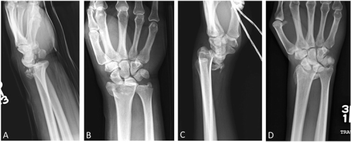
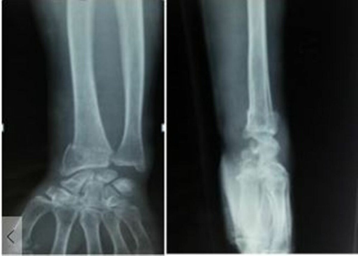
Distal radius Radius The outer shorter of the two bones of the forearm, lying parallel to the ulna and partially revolving around it. Forearm: Anatomy fractures are one of the most common fractures encountered in practice and are often associated with falling onto an outstretched hand Hand The hand constitutes the distal part of the upper limb and provides the fine, precise movements needed in activities of daily living. It consists of 5 metacarpal bones and 14 phalanges, as well as numerous muscles innervated by the median and ulnar nerves. Hand: Anatomy. These fractures are most frequently seen in older individuals, especially women. In this population, these fractures are related to an increase in falls due to gait instability Gait Instability Lateral Medullary Syndrome (Wallenberg Syndrome) with aging and associated osteoporosis Osteoporosis Osteoporosis refers to a decrease in bone mass and density leading to an increased number of fractures. There are 2 forms of osteoporosis: primary, which is commonly postmenopausal or senile; and secondary, which is a manifestation of immobilization, underlying medical disorders, or long-term use of certain medications. Osteoporosis. In younger individuals, distal radius Radius The outer shorter of the two bones of the forearm, lying parallel to the ulna and partially revolving around it. Forearm: Anatomy fractures are usually related to high-energy trauma. Individuals often present with pain Pain An unpleasant sensation induced by noxious stimuli which are detected by nerve endings of nociceptive neurons. Pain: Types and Pathways and a dinner fork deformity Deformity Examination of the Upper Limbs of the distal forearm Forearm The forearm is the region of the upper limb between the elbow and the wrist. The term "forearm" is used in anatomy to distinguish this area from the arm, a term that is commonly used to describe the entire upper limb. The forearm consists of 2 long bones (the radius and the ulna), the interosseous membrane, and multiple arteries, nerves, and muscles. Forearm: Anatomy. Diagnosis is clinical and confirmed with x-rays X-rays X-rays are high-energy particles of electromagnetic radiation used in the medical field for the generation of anatomical images. X-rays are projected through the body of a patient and onto a film, and this technique is called conventional or projectional radiography. X-rays of the wrist. Treatment can be operative or nonoperative depending on the age of the individual, articular involvement, and degree of displacement Displacement The process by which an emotional or behavioral response that is appropriate for one situation appears in another situation for which it is inappropriate. Defense Mechanisms or angulation Angulation Buckle or Torus Fracture.

Last updated: Jan 15, 2024
Editorial responsibility: Stanley Oiseth, Lindsay Jones, Evelin Maza

Advertisement

Advertisement

Advertisement

Advertisement

Advertisement

Advertisement

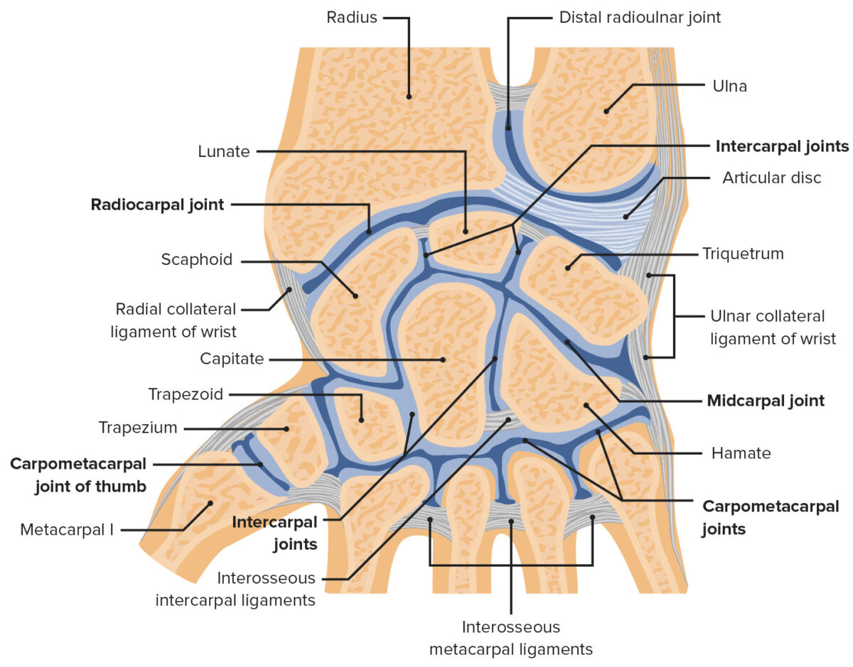
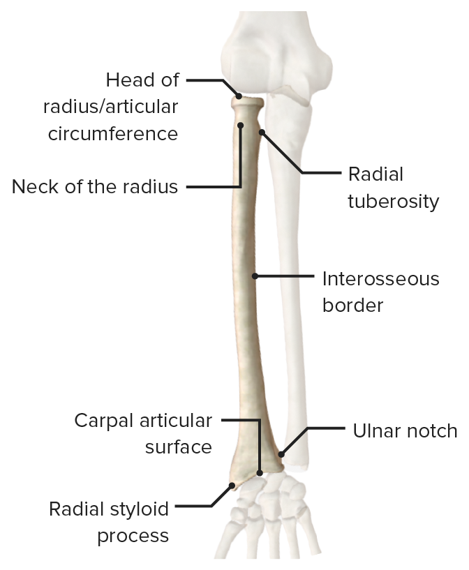
Distal Radius Anatomy

  • The radius Radius The outer shorter of the two bones of the forearm, lying parallel to the ulna and partially revolving around it. Forearm: Anatomy is the lateral bone Bone Bone is a compact type of hardened connective tissue composed of bone cells, membranes, an extracellular mineralized matrix, and central bone marrow. The 2 primary types of bone are compact and spongy. Bones: Structure and Types of the forearm Forearm The forearm is the region of the upper limb between the elbow and the wrist. The term “forearm” is used in anatomy to distinguish this area from the arm, a term that is commonly used to describe the entire upper limb. The forearm consists of 2 long bones (the radius and the ulna), the interosseous membrane, and multiple arteries, nerves, and muscles. Forearm: Anatomy. This bone Bone Bone is a compact type of hardened connective tissue composed of bone cells, membranes, an extracellular mineralized matrix, and central bone marrow. The 2 primary types of bone are compact and spongy. Bones: Structure and Types is responsible for the vast majority of the axial Axial Computed Tomography (CT) load.
  • The distal radius Radius The outer shorter of the two bones of the forearm, lying parallel to the ulna and partially revolving around it. Forearm: Anatomy:
  • The DRUJ:
    • Is primarily responsible for pronation Pronation Applies to movements of the forearm in turning the palm backward or downward. When referring to the foot, a combination of eversion and abduction movements in the tarsal and metatarsal joints (turning the foot up and in toward the midline of the body). Examination of the Upper Limbs and supination Supination Applies to movements of the forearm in turning the palm forward or upward. When referring to the foot, a combination of adduction and inversion movements of the foot. Examination of the Upper Limbs of the wrist
    • Contains a ligamentous complex, the triangular fibrocartilage Fibrocartilage A type of cartilage whose matrix contains large bundles of collagen type I. Fibrocartilage is typically found in the intervertebral disk; pubic symphysis; tibial menisci; and articular discs in synovial joints. Cartilage: Histology complex (TFCC), which may be injured in association with a distal radius Radius The outer shorter of the two bones of the forearm, lying parallel to the ulna and partially revolving around it. Forearm: Anatomy fracture Fracture A fracture is a disruption of the cortex of any bone and periosteum and is commonly due to mechanical stress after an injury or accident. Open fractures due to trauma can be a medical emergency. Fractures are frequently associated with automobile accidents, workplace injuries, and trauma. Overview of Bone Fractures

Related videos

Overview

Epidemiology

  • Prevalence Prevalence The total number of cases of a given disease in a specified population at a designated time. It is differentiated from incidence, which refers to the number of new cases in the population at a given time. Measures of Disease Frequency:
    • Distal radius Radius The outer shorter of the two bones of the forearm, lying parallel to the ulna and partially revolving around it. Forearm: Anatomy fractures: 17.5% of all fractures in adults
    • Hand Hand The hand constitutes the distal part of the upper limb and provides the fine, precise movements needed in activities of daily living. It consists of 5 metacarpal bones and 14 phalanges, as well as numerous muscles innervated by the median and ulnar nerves. Hand: Anatomy and forearm Forearm The forearm is the region of the upper limb between the elbow and the wrist. The term “forearm” is used in anatomy to distinguish this area from the arm, a term that is commonly used to describe the entire upper limb. The forearm consists of 2 long bones (the radius and the ulna), the interosseous membrane, and multiple arteries, nerves, and muscles. Forearm: Anatomy fractures account for 1.5% of ED visits.
    • Radius Radius The outer shorter of the two bones of the forearm, lying parallel to the ulna and partially revolving around it. Forearm: Anatomy and/or ulna Ulna The inner and longer bone of the forearm. Forearm: Anatomy fractures: 44% of hand Hand The hand constitutes the distal part of the upper limb and provides the fine, precise movements needed in activities of daily living. It consists of 5 metacarpal bones and 14 phalanges, as well as numerous muscles innervated by the median and ulnar nerves. Hand: Anatomy and forearm Forearm The forearm is the region of the upper limb between the elbow and the wrist. The term “forearm” is used in anatomy to distinguish this area from the arm, a term that is commonly used to describe the entire upper limb. The forearm consists of 2 long bones (the radius and the ulna), the interosseous membrane, and multiple arteries, nerves, and muscles. Forearm: Anatomy fractures
  • Characteristics:
    • Ages of individuals who most commonly present:
      • Middle-aged to elderly due to low-energy mechanisms (fall on outstretched hand Hand The hand constitutes the distal part of the upper limb and provides the fine, precise movements needed in activities of daily living. It consists of 5 metacarpal bones and 14 phalanges, as well as numerous muscles innervated by the median and ulnar nerves. Hand: Anatomy) as a result of increased bone Bone Bone is a compact type of hardened connective tissue composed of bone cells, membranes, an extracellular mineralized matrix, and central bone marrow. The 2 primary types of bone are compact and spongy. Bones: Structure and Types fragility
      • Young individuals due to high-energy mechanisms (e.g., car accidents, sports injury)
    • More common in females (F:M ratio is 2–3:1)

Etiology

  • In the elderly: fall on outstretched hand Hand The hand constitutes the distal part of the upper limb and provides the fine, precise movements needed in activities of daily living. It consists of 5 metacarpal bones and 14 phalanges, as well as numerous muscles innervated by the median and ulnar nerves. Hand: Anatomy is the most common mechanism.
  • In younger individuals:
    • High-energy trauma directly to the bone Bone Bone is a compact type of hardened connective tissue composed of bone cells, membranes, an extracellular mineralized matrix, and central bone marrow. The 2 primary types of bone are compact and spongy. Bones: Structure and Types 
    • Compression Compression Blunt Chest Trauma load driving the scaphoid or lunate into the distal radius Radius The outer shorter of the two bones of the forearm, lying parallel to the ulna and partially revolving around it. Forearm: Anatomy (e.g., punching a hard surface)

Related videos

Clinical Presentation and Diagnosis

Clinical presentation

History:

  • Individual often reports:
    • Falling on an outstretched hand Hand The hand constitutes the distal part of the upper limb and provides the fine, precise movements needed in activities of daily living. It consists of 5 metacarpal bones and 14 phalanges, as well as numerous muscles innervated by the median and ulnar nerves. Hand: Anatomy 
    • Wrist pain Pain An unpleasant sensation induced by noxious stimuli which are detected by nerve endings of nociceptive neurons. Pain: Types and Pathways with associated swelling Swelling Inflammation and deformity Deformity Examination of the Upper Limbs
  • Assess for previous fragility fractures Fragility fractures Pelvic fractures are a disruption in the cortex of a pelvic bone involving iliac wing fractures, acetabular fractures, or those causing loss of integrity of the pelvic ring (the sacrum and the 2 innominate bones). Patients often present with a history of trauma or a fall, limb length discrepancy, intense pain on palpation, and mechanical instability. Pelvic Fractures as well as the degree of energy involved in the injury.

Physical exam:

  • Evaluate for deformity Deformity Examination of the Upper Limbs and skin Skin The skin, also referred to as the integumentary system, is the largest organ of the body. The skin is primarily composed of the epidermis (outer layer) and dermis (deep layer). The epidermis is primarily composed of keratinocytes that undergo rapid turnover, while the dermis contains dense layers of connective tissue. Skin: Structure and Functions integrity:
  • Evaluation of the wrist:
    • Range of motion Range of motion The distance and direction to which a bone joint can be extended. Range of motion is a function of the condition of the joints, muscles, and connective tissues involved. Joint flexibility can be improved through appropriate muscle strength exercises. Examination of the Upper Limbs: supination Supination Applies to movements of the forearm in turning the palm forward or upward. When referring to the foot, a combination of adduction and inversion movements of the foot. Examination of the Upper Limbs, pronation Pronation Applies to movements of the forearm in turning the palm backward or downward. When referring to the foot, a combination of eversion and abduction movements in the tarsal and metatarsal joints (turning the foot up and in toward the midline of the body). Examination of the Upper Limbs, flexion Flexion Examination of the Upper Limbs, and extension Extension Examination of the Upper Limbs
    • Palpation Palpation Application of fingers with light pressure to the surface of the body to determine consistency of parts beneath in physical diagnosis; includes palpation for determining the outlines of organs. Dermatologic Examination of the ulna Ulna The inner and longer bone of the forearm. Forearm: Anatomy and radial surfaces for areas of tenderness and displacement Displacement The process by which an emotional or behavioral response that is appropriate for one situation appears in another situation for which it is inappropriate. Defense Mechanisms
    • Palpation Palpation Application of fingers with light pressure to the surface of the body to determine consistency of parts beneath in physical diagnosis; includes palpation for determining the outlines of organs. Dermatologic Examination of anatomical snuffbox to evaluate for scaphoid fracture Scaphoid Fracture Hand and Wrist Pain
  • Careful neurovascular examination:
    • Radial and ulnar pulses with capillary refill 
    • Evaluation of sensory Sensory Neurons which conduct nerve impulses to the central nervous system. Nervous System: Histology and muscular function of median, ulnar, and radial nerves
    • Index and thumb sensations should always be assessed because of increased risk of injury to median nerve Median Nerve A major nerve of the upper extremity. In humans, the fibers of the median nerve originate in the lower cervical and upper thoracic spinal cord (usually C6 to T1), travel via the brachial plexus, and supply sensory and motor innervation to parts of the forearm and hand. Cubital Fossa: Anatomy with distal radius Radius The outer shorter of the two bones of the forearm, lying parallel to the ulna and partially revolving around it. Forearm: Anatomy fracture Fracture A fracture is a disruption of the cortex of any bone and periosteum and is commonly due to mechanical stress after an injury or accident. Open fractures due to trauma can be a medical emergency. Fractures are frequently associated with automobile accidents, workplace injuries, and trauma. Overview of Bone Fractures 
  • Evaluate for associated injuries:
    • Carpal fracture Fracture A fracture is a disruption of the cortex of any bone and periosteum and is commonly due to mechanical stress after an injury or accident. Open fractures due to trauma can be a medical emergency. Fractures are frequently associated with automobile accidents, workplace injuries, and trauma. Overview of Bone Fractures/dislocations
    • DRUJ injuries
    • Carpal ligament injuries
    • Digit tendon injuries
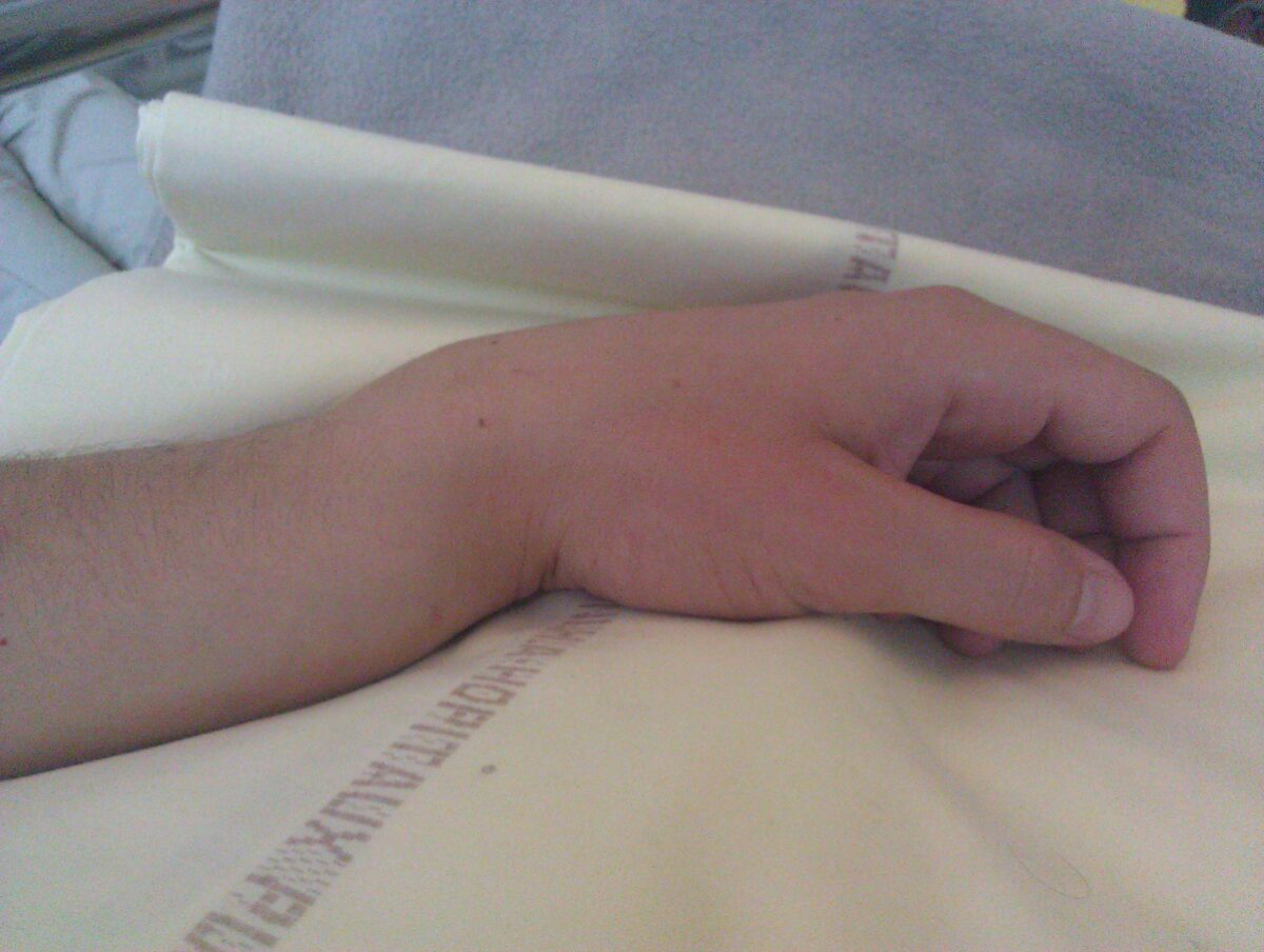
Colles fracture

Colles fracture of the left hand:
Classic dinner fork deformity with posterior displacement clearly visible.

Image: “Colles fracture of the left hand” by Sylvain Letuffe. License: Public Domain

Diagnosis

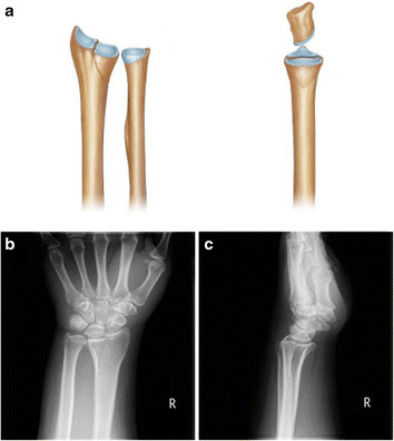
Diagnosis is clinical; however, imaging is needed for confirmation and evaluation of severity.
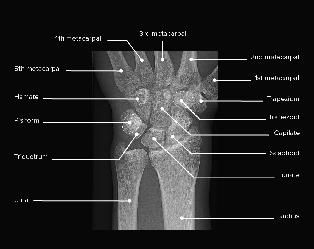
  • X-rays X-rays X-rays are high-energy particles of electromagnetic radiation used in the medical field for the generation of anatomical images. X-rays are projected through the body of a patient and onto a film, and this technique is called conventional or projectional radiography. X-rays:
    • Anteroposterior (AP) and lateral, with optional oblique
    • Evaluate six A’s:
      • Anatomy involved
      • Involvement of Articular surface
      • Alignment of the fragments
      • Degree of Angulation
      • Apex
      • Presence of Apposition
    • Acquire images of the joints above and below the injury; imaging is repeated after reduction if reduction is required.
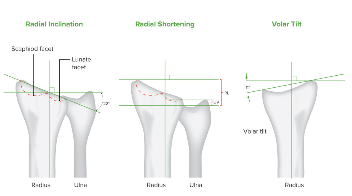
    • Findings suggestive of fracture Fracture A fracture is a disruption of the cortex of any bone and periosteum and is commonly due to mechanical stress after an injury or accident. Open fractures due to trauma can be a medical emergency. Fractures are frequently associated with automobile accidents, workplace injuries, and trauma. Overview of Bone Fractures:
      • Radial shortening of ≥ 5 mm
      • Radial inclination change of ≥ 5 degrees
      • Articular step-off of ≥ 2 mm
      • Volar tilt with dorsal angulation Angulation Buckle or Torus Fracture of ≥ 5 degrees
  • CT scan:
    • May be used to evaluate intraarticular involvement 
    • Useful for surgical planning
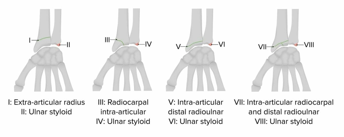
  • Frykman classification of distal radius Radius The outer shorter of the two bones of the forearm, lying parallel to the ulna and partially revolving around it. Forearm: Anatomy fractures:
Frykman classification of distal radius fractures

Frykman classification of distal radius fractures

Image by Lecturio.
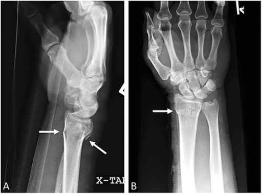
Specific examples of distal radius Radius The outer shorter of the two bones of the forearm, lying parallel to the ulna and partially revolving around it. Forearm: Anatomy fractures

  • Colles fracture Fracture A fracture is a disruption of the cortex of any bone and periosteum and is commonly due to mechanical stress after an injury or accident. Open fractures due to trauma can be a medical emergency. Fractures are frequently associated with automobile accidents, workplace injuries, and trauma. Overview of Bone Fractures:
    • A common fracture Fracture A fracture is a disruption of the cortex of any bone and periosteum and is commonly due to mechanical stress after an injury or accident. Open fractures due to trauma can be a medical emergency. Fractures are frequently associated with automobile accidents, workplace injuries, and trauma. Overview of Bone Fractures of the distal radius Radius The outer shorter of the two bones of the forearm, lying parallel to the ulna and partially revolving around it. Forearm: Anatomy, often low-energy
    • Generally extraarticular and dorsally displaced
  • Smith fracture Fracture A fracture is a disruption of the cortex of any bone and periosteum and is commonly due to mechanical stress after an injury or accident. Open fractures due to trauma can be a medical emergency. Fractures are frequently associated with automobile accidents, workplace injuries, and trauma. Overview of Bone Fractures:
    • Considered a reverse Colles fracture Fracture A fracture is a disruption of the cortex of any bone and periosteum and is commonly due to mechanical stress after an injury or accident. Open fractures due to trauma can be a medical emergency. Fractures are frequently associated with automobile accidents, workplace injuries, and trauma. Overview of Bone Fractures 
    • Distal radius Radius The outer shorter of the two bones of the forearm, lying parallel to the ulna and partially revolving around it. Forearm: Anatomy angulation Angulation Buckle or Torus Fracture is in the palmar direction.
    • Often seen if the fall occurs on a flexed wrist
  • Die-punch fracture Fracture A fracture is a disruption of the cortex of any bone and periosteum and is commonly due to mechanical stress after an injury or accident. Open fractures due to trauma can be a medical emergency. Fractures are frequently associated with automobile accidents, workplace injuries, and trauma. Overview of Bone Fractures:
    • Fracture Fracture A fracture is a disruption of the cortex of any bone and periosteum and is commonly due to mechanical stress after an injury or accident. Open fractures due to trauma can be a medical emergency. Fractures are frequently associated with automobile accidents, workplace injuries, and trauma. Overview of Bone Fractures of the distal radius Radius The outer shorter of the two bones of the forearm, lying parallel to the ulna and partially revolving around it. Forearm: Anatomy resulting from an axial Axial Computed Tomography (CT) loading force
    • Intraarticular fracture Intraarticular fracture Fractures of the articular surface of a bone. Overview of Bone Fractures of the lunate fossa of the distal radius Radius The outer shorter of the two bones of the forearm, lying parallel to the ulna and partially revolving around it. Forearm: Anatomy
  • Barton fracture Fracture A fracture is a disruption of the cortex of any bone and periosteum and is commonly due to mechanical stress after an injury or accident. Open fractures due to trauma can be a medical emergency. Fractures are frequently associated with automobile accidents, workplace injuries, and trauma. Overview of Bone Fractures:
    • Intraarticular fracture Intraarticular fracture Fractures of the articular surface of a bone. Overview of Bone Fractures/ subluxation Subluxation Radial Head Subluxation (Nursemaid’s Elbow) involving the dorsal or volar rim of the distal radius Radius The outer shorter of the two bones of the forearm, lying parallel to the ulna and partially revolving around it. Forearm: Anatomy
    • Often appears as a triangular fragment of the distal radial styloid on x-ray X-ray Penetrating electromagnetic radiation emitted when the inner orbital electrons of an atom are excited and release radiant energy. X-ray wavelengths range from 1 pm to 10 nm. Hard x-rays are the higher energy, shorter wavelength x-rays. Soft x-rays or grenz rays are less energetic and longer in wavelength. The short wavelength end of the x-ray spectrum overlaps the gamma rays wavelength range. The distinction between gamma rays and x-rays is based on their radiation source. Pulmonary Function Tests
  • Hutchinson fracture Fracture A fracture is a disruption of the cortex of any bone and periosteum and is commonly due to mechanical stress after an injury or accident. Open fractures due to trauma can be a medical emergency. Fractures are frequently associated with automobile accidents, workplace injuries, and trauma. Overview of Bone Fractures (chauffeur’s fracture Fracture A fracture is a disruption of the cortex of any bone and periosteum and is commonly due to mechanical stress after an injury or accident. Open fractures due to trauma can be a medical emergency. Fractures are frequently associated with automobile accidents, workplace injuries, and trauma. Overview of Bone Fractures):
    • Fracture Fracture A fracture is a disruption of the cortex of any bone and periosteum and is commonly due to mechanical stress after an injury or accident. Open fractures due to trauma can be a medical emergency. Fractures are frequently associated with automobile accidents, workplace injuries, and trauma. Overview of Bone Fractures through the base of the radial styloid
    • Occurs with forced hyperextension of the wrist

Management

Management, whether nonsurgical or surgical, is based on the type of fracture Fracture A fracture is a disruption of the cortex of any bone and periosteum and is commonly due to mechanical stress after an injury or accident. Open fractures due to trauma can be a medical emergency. Fractures are frequently associated with automobile accidents, workplace injuries, and trauma. Overview of Bone Fractures (based on articular involvement and displacement Displacement The process by which an emotional or behavioral response that is appropriate for one situation appears in another situation for which it is inappropriate. Defense Mechanisms) and the age and activity level of the individual.
Nonsurgical management

  • Splinting:
    • Often used initially to allow swelling Swelling Inflammation to decrease before casting
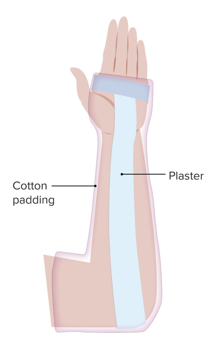
    • Simple splint (volar or sugar tong style) can be done for fractures with minimal/no displacement Displacement The process by which an emotional or behavioral response that is appropriate for one situation appears in another situation for which it is inappropriate. Defense Mechanisms or after an adequate closed reduction Closed Reduction Radial Head Subluxation (Nursemaid’s Elbow) ( x-rays X-rays X-rays are high-energy particles of electromagnetic radiation used in the medical field for the generation of anatomical images. X-rays are projected through the body of a patient and onto a film, and this technique is called conventional or projectional radiography. X-rays before and after reduction should be assessed) 
    • In a sugar tong splint, the elbow is flexed to 90 degrees, with neutral positioning of the wrist
  • Swelling Swelling Inflammation and pain Pain An unpleasant sensation induced by noxious stimuli which are detected by nerve endings of nociceptive neurons. Pain: Types and Pathways management:
    • Circumferential casting should not be done in the acute setting because continued swelling Swelling Inflammation may contribute to neurovascular injury (distal ischemia Ischemia A hypoperfusion of the blood through an organ or tissue caused by a pathologic constriction or obstruction of its blood vessels, or an absence of blood circulation. Ischemic Cell Damage and carpal tunnel Carpal Tunnel The carpal tunnel is formed by the transverse carpal ligament (flexor retinaculum) superiorly and the carpal bones inferiorly. Carpal Tunnel Syndrome syndrome most common).
    • Elevation and ice are helpful to reduce swelling Swelling Inflammation and to assist in pain Pain An unpleasant sensation induced by noxious stimuli which are detected by nerve endings of nociceptive neurons. Pain: Types and Pathways control for a properly reduced or nondisplaced fracture Fracture A fracture is a disruption of the cortex of any bone and periosteum and is commonly due to mechanical stress after an injury or accident. Open fractures due to trauma can be a medical emergency. Fractures are frequently associated with automobile accidents, workplace injuries, and trauma. Overview of Bone Fractures.
    • Active range of motion Range of motion The distance and direction to which a bone joint can be extended. Range of motion is a function of the condition of the joints, muscles, and connective tissues involved. Joint flexibility can be improved through appropriate muscle strength exercises. Examination of the Upper Limbs of shoulder and fingers are allowed and encouraged.
    • Pain Pain An unpleasant sensation induced by noxious stimuli which are detected by nerve endings of nociceptive neurons. Pain: Types and Pathways management should be tailored to the individual’s needs, using the smallest amount of the least potent medication possible.
Forearm sugar tong splint

Forearm sugar tong splint:
A simple splint, the sugar tong splint is indicated in distal radius and ulnar fractures that are not displaced. The splint impedes pronation/supination and immobilizes the elbow joint.

Image by Lecturio.

Surgical management

Surgical management is indicated in the presence of:

  • Significant displacement Displacement The process by which an emotional or behavioral response that is appropriate for one situation appears in another situation for which it is inappropriate. Defense Mechanisms or substantial articular involvement as demonstrated by:
    • Intraarticular step-off of the distal radius Radius The outer shorter of the two bones of the forearm, lying parallel to the ulna and partially revolving around it. Forearm: Anatomy
    • Increased dorsal tilt
    • Loss of radial length
  • Neurovascular compromise without resolution after reduction
  • Inadequate reduction
  • Associated instability

Prognosis Prognosis A prediction of the probable outcome of a disease based on a individual’s condition and the usual course of the disease as seen in similar situations. Non-Hodgkin Lymphomas

  • Prognosis Prognosis A prediction of the probable outcome of a disease based on a individual’s condition and the usual course of the disease as seen in similar situations. Non-Hodgkin Lymphomas is favorable.
  • The majority of individuals resume their previous level of activity.
  • Some individuals have resultant loss of motion, instability, and posttraumatic degenerative joint disease.

Differential Diagnosis

  • Scaphoid fractures: A fall on an outstretched hand Hand The hand constitutes the distal part of the upper limb and provides the fine, precise movements needed in activities of daily living. It consists of 5 metacarpal bones and 14 phalanges, as well as numerous muscles innervated by the median and ulnar nerves. Hand: Anatomy is associated with carpal bone Bone Bone is a compact type of hardened connective tissue composed of bone cells, membranes, an extracellular mineralized matrix, and central bone marrow. The 2 primary types of bone are compact and spongy. Bones: Structure and Types injuries. Physicians Physicians Individuals licensed to practice medicine. Clinician–Patient Relationship should specifically evaluate for scaphoid fractures by palpating the anatomical snuffbox area of the wrist. Range of motion Range of motion The distance and direction to which a bone joint can be extended. Range of motion is a function of the condition of the joints, muscles, and connective tissues involved. Joint flexibility can be improved through appropriate muscle strength exercises. Examination of the Upper Limbs is usually not restricted, and swelling Swelling Inflammation/ deformity Deformity Examination of the Upper Limbs of the wrist are usually minimal. Diagnosis is clinical, combined with radiographic findings. Treatment varies based on severity, ranging from surgical fixation to immobilization Immobilization Delirium in a short-arm thumb spica cast with the wrist in slight extension Extension Examination of the Upper Limbs for 4–6 weeks.
  • Triangular fibrocartilage Fibrocartilage A type of cartilage whose matrix contains large bundles of collagen type I. Fibrocartilage is typically found in the intervertebral disk; pubic symphysis; tibial menisci; and articular discs in synovial joints. Cartilage: Histology complex injury: ligamentous joint that stabilizes the radioulnar juncture and commonly injured with falls on an outstretched hand Falls On An Outstretched Hand Greenstick Fracture. Symptoms include pain Pain An unpleasant sensation induced by noxious stimuli which are detected by nerve endings of nociceptive neurons. Pain: Types and Pathways over the ulnar aspect of the wrist, increasing with forearm Forearm The forearm is the region of the upper limb between the elbow and the wrist. The term “forearm” is used in anatomy to distinguish this area from the arm, a term that is commonly used to describe the entire upper limb. The forearm consists of 2 long bones (the radius and the ulna), the interosseous membrane, and multiple arteries, nerves, and muscles. Forearm: Anatomy rotation Rotation Motion of an object in which either one or more points on a line are fixed. It is also the motion of a particle about a fixed point. X-rays and ulnar deviation. Management is primarily nonsurgical, involving NSAIDs NSAIDS Primary vs Secondary Headaches, ice, and splinting.
  • Wrist sprain: common term for a strain of the radiocarpal Radiocarpal Wrist Joint: Anatomy ligaments. Presentation is typically with acute wrist pain Pain An unpleasant sensation induced by noxious stimuli which are detected by nerve endings of nociceptive neurons. Pain: Types and Pathways with minimal restriction of movement, usually after trauma. Management is conservative and supportive only. A sprained wrist can only be a diagnosis of exclusion, after all other pathologic processes have been ruled out.
  • Carpal fractures: common injuries that often occur secondary to falls. Fractures of the capitate, lunate, and trapezoid usually present with swelling Swelling Inflammation, tenderness, and reduced range of motion Range of motion The distance and direction to which a bone joint can be extended. Range of motion is a function of the condition of the joints, muscles, and connective tissues involved. Joint flexibility can be improved through appropriate muscle strength exercises. Examination of the Upper Limbs of the wrist. Surgical repair may be indicated, depending on which bone Bone Bone is a compact type of hardened connective tissue composed of bone cells, membranes, an extracellular mineralized matrix, and central bone marrow. The 2 primary types of bone are compact and spongy. Bones: Structure and Types is fractured.

References

  1. Meena, S., Sharma, P., Sambharia, A.K., Dawar, A. (2014). Fractures of distal radius: an overview. J Family Med Prim Care 3:325–332. https://pubmed.ncbi.nlm.nih.gov/25657938/ 
  2. Chung, K.C., Spilson, S.V. (2001). The frequency and epidemiology of hand and forearm fractures in the United States. J Hand Surg Am 26:908–915. https://pubmed.ncbi.nlm.nih.gov/11561245/ 
  3. Lawson, G.M., Hajducka, C., McQueen, M.M. (1995). Sports fractures of the distal radius—epidemiology and outcome. Injury 26:33–36. https://pubmed.ncbi.nlm.nih.gov/7868207/ 
  4. O’Neill, T.W., UK Colles’ Fracture Study Group, et al. (2001). Incidence of distal forearm fracture in British men and women. Osteoporos Int 12:555–558. https://pubmed.ncbi.nlm.nih.gov/11527052/ 
  5. Gutow, A.P. (2005). Avoidance and treatment of complications of distal radius fractures. Hand Clin 21:295–305. https://pubmed.ncbi.nlm.nih.gov/16039441/ 
  6. Bozentka, D.J., et al. (2002). Digital radiographs in the assessment of distal radius fracture parameters. Clin Orthop Relat Res 397:409–413. https://pubmed.ncbi.nlm.nih.gov/11953635/ 
  7. Lafontaine, M., Hardy, D., Delince, P. (1989). Stability assessment of distal radius fractures. Injury 20:208–210. https://pubmed.ncbi.nlm.nih.gov/2592094/

Fall in Love
with Lecturio

🍁
Get 30% OFF all premium plans!
Copy your code Copied!

Create your free account or log in to continue reading!

Sign up now and get free access to Lecturio with concept pages, medical videos, and questions for your medical education.

User Reviews

Details