Advertisement

Advertisement

Advertisement
Advertisement

Orbit and Extraocular Muscles: Anatomy

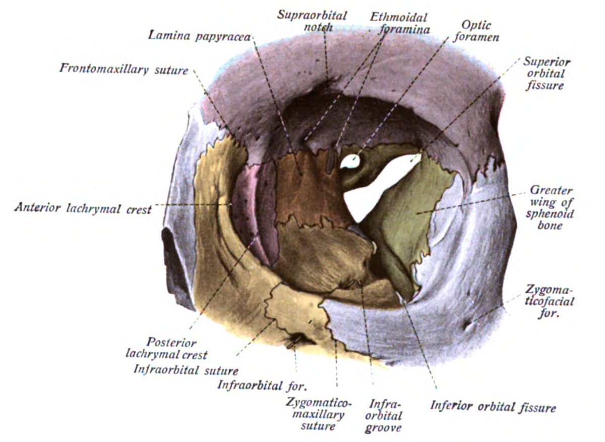
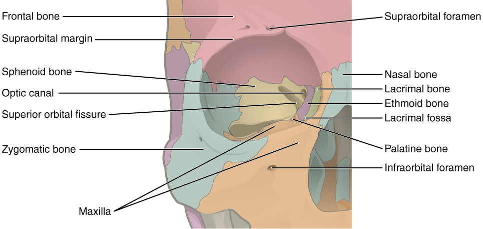
The orbit is the cavity of the skull Skull The skull (cranium) is the skeletal structure of the head supporting the face and forming a protective cavity for the brain. The skull consists of 22 bones divided into the viscerocranium (facial skeleton) and the neurocranium. Skull: Anatomy in which the eye and its appendages are situated. The orbit is composed of 7 bones and has a pyramidal shape, with its apex pointed posteromedially. The orbital contents comprise the eye; orbital and retrobulbar fascia Fascia Layers of connective tissue of variable thickness. The superficial fascia is found immediately below the skin; the deep fascia invests muscles, nerves, and other organs. Cellulitis; extraocular muscles; cranial nerves Cranial nerves There are 12 pairs of cranial nerves (CNs), which run from the brain to various parts of the head, neck, and trunk. The CNs can be sensory or motor or both. The CNs are named and numbered in Roman numerals according to their location, from the front to the back of the brain. The 12 Cranial Nerves: Overview and Functions II, III, IV, V, and VI; blood vessels; fat; lacrimal gland with its sac and nasolacrimal duct Nasolacrimal Duct Nasolacrimal duct. Dacryocystitis; eyelids Eyelids Each of the upper and lower folds of skin which cover the eye when closed. Blepharitis; palpebral and suspensory ligaments; ciliary ganglion Ciliary Ganglion Pupil: Physiology and Abnormalities; and short ciliary nerves.

Last updated: Dec 5, 2024

Editorial responsibility: Stanley Oiseth, Lindsay Jones, Evelin Maza

Advertisement

Advertisement

Advertisement

Advertisement

Advertisement

Advertisement

Bones and Structure of the Orbit

  • Shaped like a quadrangular pyramidal cavern in the upper face, with a superficial base (orbital margin) and a deep, posteromedial apex
    • Orbital margin: 
      • Superior margin: frontal Frontal The bone that forms the frontal aspect of the skull. Its flat part forms the forehead, articulating inferiorly with the nasal bone and the cheek bone on each side of the face. Skull: Anatomy bone Bone Bone is a compact type of hardened connective tissue composed of bone cells, membranes, an extracellular mineralized matrix, and central bone marrow. The 2 primary types of bone are compact and spongy. Bones: Structure and Types 
      • Medial margin: frontal Frontal The bone that forms the frontal aspect of the skull. Its flat part forms the forehead, articulating inferiorly with the nasal bone and the cheek bone on each side of the face. Skull: Anatomy process of the maxilla Maxilla One of a pair of irregularly shaped bones that form the upper jaw. A maxillary bone provides tooth sockets for the superior teeth, forms part of the orbit, and contains the maxillary sinus. Skull: Anatomy
      • Inferior margin: zygomatic Zygomatic Either of a pair of bones that form the prominent part of the cheek and contribute to the orbit on each side of the skull. Skull: Anatomy process of the maxilla Maxilla One of a pair of irregularly shaped bones that form the upper jaw. A maxillary bone provides tooth sockets for the superior teeth, forms part of the orbit, and contains the maxillary sinus. Skull: Anatomy and zygomatic Zygomatic Either of a pair of bones that form the prominent part of the cheek and contribute to the orbit on each side of the skull. Skull: Anatomy bone Bone Bone is a compact type of hardened connective tissue composed of bone cells, membranes, an extracellular mineralized matrix, and central bone marrow. The 2 primary types of bone are compact and spongy. Bones: Structure and Types
      • Lateral margin: zygomatic Zygomatic Either of a pair of bones that form the prominent part of the cheek and contribute to the orbit on each side of the skull. Skull: Anatomy process of the frontal Frontal The bone that forms the frontal aspect of the skull. Its flat part forms the forehead, articulating inferiorly with the nasal bone and the cheek bone on each side of the face. Skull: Anatomy bone Bone Bone is a compact type of hardened connective tissue composed of bone cells, membranes, an extracellular mineralized matrix, and central bone marrow. The 2 primary types of bone are compact and spongy. Bones: Structure and Types and frontal Frontal The bone that forms the frontal aspect of the skull. Its flat part forms the forehead, articulating inferiorly with the nasal bone and the cheek bone on each side of the face. Skull: Anatomy process of the zygomatic Zygomatic Either of a pair of bones that form the prominent part of the cheek and contribute to the orbit on each side of the skull. Skull: Anatomy bone Bone Bone is a compact type of hardened connective tissue composed of bone cells, membranes, an extracellular mineralized matrix, and central bone marrow. The 2 primary types of bone are compact and spongy. Bones: Structure and Types
    • Apex: optic foramen
  • Walls: covered with periosteum Periosteum Thin outer membrane that surrounds a bone. It contains connective tissue, capillaries, nerves, and a number of cell types. Bones: Structure and Types (periorbita)
    • Superior (roof): orbital part of the frontal Frontal The bone that forms the frontal aspect of the skull. Its flat part forms the forehead, articulating inferiorly with the nasal bone and the cheek bone on each side of the face. Skull: Anatomy and lesser wing of the sphenoid
    • Medial: orbital plate of ethmoid bone Bone Bone is a compact type of hardened connective tissue composed of bone cells, membranes, an extracellular mineralized matrix, and central bone marrow. The 2 primary types of bone are compact and spongy. Bones: Structure and Types, body of sphenoid bone Bone Bone is a compact type of hardened connective tissue composed of bone cells, membranes, an extracellular mineralized matrix, and central bone marrow. The 2 primary types of bone are compact and spongy. Bones: Structure and Types, frontal Frontal The bone that forms the frontal aspect of the skull. Its flat part forms the forehead, articulating inferiorly with the nasal bone and the cheek bone on each side of the face. Skull: Anatomy bone Bone Bone is a compact type of hardened connective tissue composed of bone cells, membranes, an extracellular mineralized matrix, and central bone marrow. The 2 primary types of bone are compact and spongy. Bones: Structure and Types, lacrimal bone Bone Bone is a compact type of hardened connective tissue composed of bone cells, membranes, an extracellular mineralized matrix, and central bone marrow. The 2 primary types of bone are compact and spongy. Bones: Structure and Types, and maxilla Maxilla One of a pair of irregularly shaped bones that form the upper jaw. A maxillary bone provides tooth sockets for the superior teeth, forms part of the orbit, and contains the maxillary sinus. Skull: Anatomy
    • Inferior (floor): orbital surface of the maxilla Maxilla One of a pair of irregularly shaped bones that form the upper jaw. A maxillary bone provides tooth sockets for the superior teeth, forms part of the orbit, and contains the maxillary sinus. Skull: Anatomy, zygomatic Zygomatic Either of a pair of bones that form the prominent part of the cheek and contribute to the orbit on each side of the skull. Skull: Anatomy bone Bone Bone is a compact type of hardened connective tissue composed of bone cells, membranes, an extracellular mineralized matrix, and central bone marrow. The 2 primary types of bone are compact and spongy. Bones: Structure and Types, and palatine bone Bone Bone is a compact type of hardened connective tissue composed of bone cells, membranes, an extracellular mineralized matrix, and central bone marrow. The 2 primary types of bone are compact and spongy. Bones: Structure and Types
    • Lateral: greater wing of sphenoid, orbital plate of frontal Frontal The bone that forms the frontal aspect of the skull. Its flat part forms the forehead, articulating inferiorly with the nasal bone and the cheek bone on each side of the face. Skull: Anatomy bone Bone Bone is a compact type of hardened connective tissue composed of bone cells, membranes, an extracellular mineralized matrix, and central bone marrow. The 2 primary types of bone are compact and spongy. Bones: Structure and Types, and frontal Frontal The bone that forms the frontal aspect of the skull. Its flat part forms the forehead, articulating inferiorly with the nasal bone and the cheek bone on each side of the face. Skull: Anatomy process of zygomatic Zygomatic Either of a pair of bones that form the prominent part of the cheek and contribute to the orbit on each side of the skull. Skull: Anatomy bone Bone Bone is a compact type of hardened connective tissue composed of bone cells, membranes, an extracellular mineralized matrix, and central bone marrow. The 2 primary types of bone are compact and spongy. Bones: Structure and Types
  • Important openings: optic foramen or canal, anterior and posterior ethmoidal foramina, superior and inferior orbital fissures, infraorbital groove, and supraorbital notch
Location Contents
Optic foramen or canal Apex, bordered by the body and lesser wing of the sphenoid
  • Optic nerve Optic nerve The 2nd cranial nerve which conveys visual information from the retina to the brain. The nerve carries the axons of the retinal ganglion cells which sort at the optic chiasm and continue via the optic tracts to the brain. The largest projection is to the lateral geniculate nuclei; other targets include the superior colliculi and the suprachiasmatic nuclei. Though known as the second cranial nerve, it is considered part of the central nervous system. The 12 Cranial Nerves: Overview and Functions (CN II)
  • Ophthalmic artery Ophthalmic artery Artery originating from the internal carotid artery and distributing to the eye, orbit and adjacent facial structures. Eye: Anatomy
Ethmoidal foramina
  • Junction between the superior and medial orbital walls
  • In the ethmoid bone Bone Bone is a compact type of hardened connective tissue composed of bone cells, membranes, an extracellular mineralized matrix, and central bone marrow. The 2 primary types of bone are compact and spongy. Bones: Structure and Types, lateral to olfactory groove
Anterior and posterior ethmoidal veins Veins Veins are tubular collections of cells, which transport deoxygenated blood and waste from the capillary beds back to the heart. Veins are classified into 3 types: small veins/venules, medium veins, and large veins. Each type contains 3 primary layers: tunica intima, tunica media, and tunica adventitia. Veins: Histology, arteries Arteries Arteries are tubular collections of cells that transport oxygenated blood and nutrients from the heart to the tissues of the body. The blood passes through the arteries in order of decreasing luminal diameter, starting in the largest artery (the aorta) and ending in the small arterioles. Arteries are classified into 3 types: large elastic arteries, medium muscular arteries, and small arteries and arterioles. Arteries: Histology, and nerves
Superior orbital fissure Fissure A crack or split that extends into the dermis Generalized and Localized Rashes Between the greater and lesser wings of the sphenoid bone Bone Bone is a compact type of hardened connective tissue composed of bone cells, membranes, an extracellular mineralized matrix, and central bone marrow. The 2 primary types of bone are compact and spongy. Bones: Structure and Types Inside the common tendinous ring:
  • Nasociliary nerve
  • Superior and inferior branches of the oculomotor nerve Oculomotor nerve The 3D cranial nerve. The oculomotor nerve sends motor fibers to the levator muscles of the eyelid and to the superior rectus, inferior rectus, and inferior oblique muscles of the eye. It also sends parasympathetic efferents (via the ciliary ganglion) to the muscles controlling pupillary constriction and accommodation. The motor fibers originate in the oculomotor nuclei of the midbrain. The 12 Cranial Nerves: Overview and Functions (CN III)
  • Abducens (CN VI) nerve
Outside the common tendinous ring:
  • Frontal Frontal The bone that forms the frontal aspect of the skull. Its flat part forms the forehead, articulating inferiorly with the nasal bone and the cheek bone on each side of the face. Skull: Anatomy nerve (branch off ophthalmic division of CN V)
  • Lacrimal nerve
  • Trochlear (CN IV) nerve
  • Superior ophthalmic vein
Inferior orbital fissure Fissure A crack or split that extends into the dermis Generalized and Localized Rashes
  • Lateral border of orbital floor Orbital Floor Orbital Fractures
  • Formed by greater wing of sphenoid superiorly and palatine and maxillary bones inferiorly
  • Inferior ophthalmic vein
  • Infraorbital artery and vein
  • Branches of the maxillary division of the trigeminal nerve Trigeminal nerve The 5th and largest cranial nerve. The trigeminal nerve is a mixed motor and sensory nerve. The larger sensory part forms the ophthalmic, mandibular, and maxillary nerves which carry afferents sensitive to external or internal stimuli from the skin, muscles, and joints of the face and mouth and from the teeth. Most of these fibers originate from cells of the trigeminal ganglion and project to the trigeminal nucleus of the brain stem. The smaller motor part arises from the brain stem trigeminal motor nucleus and innervates the muscles of mastication. The 12 Cranial Nerves: Overview and Functions (CN V2)— zygomatic Zygomatic Either of a pair of bones that form the prominent part of the cheek and contribute to the orbit on each side of the skull. Skull: Anatomy and infraorbital nerves
  • Orbital branches of the pterygopalatine ganglion
Infraorbital foramen Middle of the orbital floor Orbital Floor Orbital Fractures ( maxilla Maxilla One of a pair of irregularly shaped bones that form the upper jaw. A maxillary bone provides tooth sockets for the superior teeth, forms part of the orbit, and contains the maxillary sinus. Skull: Anatomy) Exit of the infraorbital vein, artery, and nerve
Supraorbital notch or foramen Superior margin of the orbit ( frontal Frontal The bone that forms the frontal aspect of the skull. Its flat part forms the forehead, articulating inferiorly with the nasal bone and the cheek bone on each side of the face. Skull: Anatomy bone Bone Bone is a compact type of hardened connective tissue composed of bone cells, membranes, an extracellular mineralized matrix, and central bone marrow. The 2 primary types of bone are compact and spongy. Bones: Structure and Types) Exit of the supraorbital vein, artery, and nerve

Mnemonic 1

To help memorize the bones that make up the orbit, remember: Many Friendly Zebras Enjoy Lazy Summer Picnics

  • Many: M axilla Axilla The axilla is a pyramid-shaped space located between the upper thorax and the arm. The axilla has a base, an apex, and 4 walls (anterior, medial, lateral, posterior). The base of the pyramid is made up of the axillary skin. The apex is the axillary inlet, located between the 1st rib, superior border of the scapula, and clavicle. Axilla and Brachial Plexus: Anatomy
  • Friendly: Frontal bone Bone Bone is a compact type of hardened connective tissue composed of bone cells, membranes, an extracellular mineralized matrix, and central bone marrow. The 2 primary types of bone are compact and spongy. Bones: Structure and Types
  • Zebras: Zygomatic bone Bone Bone is a compact type of hardened connective tissue composed of bone cells, membranes, an extracellular mineralized matrix, and central bone marrow. The 2 primary types of bone are compact and spongy. Bones: Structure and Types
  • Enjoy: Ethmoid bone Bone Bone is a compact type of hardened connective tissue composed of bone cells, membranes, an extracellular mineralized matrix, and central bone marrow. The 2 primary types of bone are compact and spongy. Bones: Structure and Types
  • Lazy: Lacrimal bone Bone Bone is a compact type of hardened connective tissue composed of bone cells, membranes, an extracellular mineralized matrix, and central bone marrow. The 2 primary types of bone are compact and spongy. Bones: Structure and Types
  • Summer: Sphenoid bone Bone Bone is a compact type of hardened connective tissue composed of bone cells, membranes, an extracellular mineralized matrix, and central bone marrow. The 2 primary types of bone are compact and spongy. Bones: Structure and Types
  • Picnics: Palatine bone Bone Bone is a compact type of hardened connective tissue composed of bone cells, membranes, an extracellular mineralized matrix, and central bone marrow. The 2 primary types of bone are compact and spongy. Bones: Structure and Types

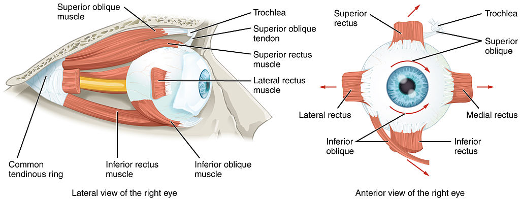
Extraocular Muscles

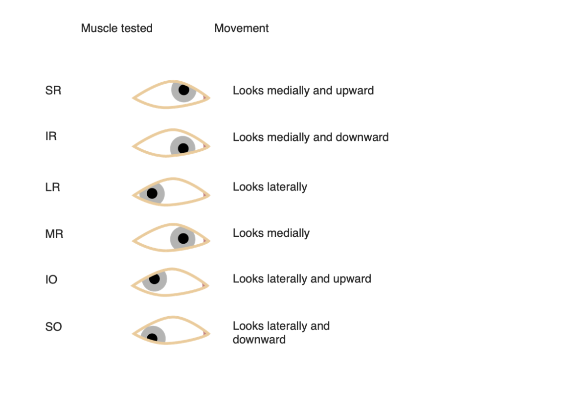
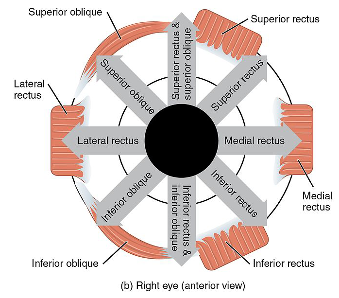
Muscle Origin Insertion Irrigation Innervation Function
Medial rectus Annulus of Zinn (common tendinous ring) Anterior, medial surface of the eye Inferior muscular branch of ophthalmic artery Ophthalmic artery Artery originating from the internal carotid artery and distributing to the eye, orbit and adjacent facial structures. Eye: Anatomy Inferior branch of oculomotor nerve Oculomotor nerve The 3D cranial nerve. The oculomotor nerve sends motor fibers to the levator muscles of the eyelid and to the superior rectus, inferior rectus, and inferior oblique muscles of the eye. It also sends parasympathetic efferents (via the ciliary ganglion) to the muscles controlling pupillary constriction and accommodation. The motor fibers originate in the oculomotor nuclei of the midbrain. The 12 Cranial Nerves: Overview and Functions (CN III) Adduction Adduction Examination of the Upper Limbs
Lateral rectus Anterior, lateral surface of the eye Lacrimal artery Abducens nerve Abducens nerve The 6th cranial nerve which originates in the abducens nucleus of the pons and sends motor fibers to the lateral rectus muscles of the eye. Damage to the nerve or its nucleus disrupts horizontal eye movement control. The 12 Cranial Nerves: Overview and Functions (CN VI) Abduction Abduction Examination of the Upper Limbs
Inferior rectus Anterior, inferior surface of the eye Inferior muscular branch of ophthalmic artery Ophthalmic artery Artery originating from the internal carotid artery and distributing to the eye, orbit and adjacent facial structures. Eye: Anatomy and the infraorbital branch of the maxillary artery Inferior branch of oculomotor nerve Oculomotor nerve The 3D cranial nerve. The oculomotor nerve sends motor fibers to the levator muscles of the eyelid and to the superior rectus, inferior rectus, and inferior oblique muscles of the eye. It also sends parasympathetic efferents (via the ciliary ganglion) to the muscles controlling pupillary constriction and accommodation. The motor fibers originate in the oculomotor nuclei of the midbrain. The 12 Cranial Nerves: Overview and Functions (CN III) Depression, extorsion, and adduction Adduction Examination of the Upper Limbs. In abduction Abduction Examination of the Upper Limbs: only depresses
Superior rectus Anterior, superior surface of the eye Superior muscular branch of ophthalmic artery Ophthalmic artery Artery originating from the internal carotid artery and distributing to the eye, orbit and adjacent facial structures. Eye: Anatomy Superior branch of the oculomotor nerve Oculomotor nerve The 3D cranial nerve. The oculomotor nerve sends motor fibers to the levator muscles of the eyelid and to the superior rectus, inferior rectus, and inferior oblique muscles of the eye. It also sends parasympathetic efferents (via the ciliary ganglion) to the muscles controlling pupillary constriction and accommodation. The motor fibers originate in the oculomotor nuclei of the midbrain. The 12 Cranial Nerves: Overview and Functions (CN III) Elevation, intorsion, and adduction Adduction Examination of the Upper Limbs. In abduction Abduction Examination of the Upper Limbs: only elevates
Superior oblique Lesser wing of sphenoid, medial to optic canal Posterior, superior, lateral surface of the eye Superior muscular branch of the ophthalmic artery Ophthalmic artery Artery originating from the internal carotid artery and distributing to the eye, orbit and adjacent facial structures. Eye: Anatomy Trochlear nerve Trochlear nerve The 4th cranial nerve. The trochlear nerve carries the motor innervation of the superior oblique muscles of the eye. The 12 Cranial Nerves: Overview and Functions (CN IV) Intorsion, depression, and abduction Abduction Examination of the Upper Limbs
Inferior oblique Lateral to the lacrimal groove ( maxilla Maxilla One of a pair of irregularly shaped bones that form the upper jaw. A maxillary bone provides tooth sockets for the superior teeth, forms part of the orbit, and contains the maxillary sinus. Skull: Anatomy) Posterior, inferior, lateral surface of the eye Inferior branch of the ophthalmic artery Ophthalmic artery Artery originating from the internal carotid artery and distributing to the eye, orbit and adjacent facial structures. Eye: Anatomy and infraorbital artery Inferior branch of oculomotor nerve Oculomotor nerve The 3D cranial nerve. The oculomotor nerve sends motor fibers to the levator muscles of the eyelid and to the superior rectus, inferior rectus, and inferior oblique muscles of the eye. It also sends parasympathetic efferents (via the ciliary ganglion) to the muscles controlling pupillary constriction and accommodation. The motor fibers originate in the oculomotor nuclei of the midbrain. The 12 Cranial Nerves: Overview and Functions (CN III) Extorsion, elevation, and abduction Abduction Examination of the Upper Limbs
Levator palpebrae superioris Lesser wing of the sphenoid, above the optic canal Tarsal plate of upper eyelid Supraorbital branch of the ophthalmic artery Ophthalmic artery Artery originating from the internal carotid artery and distributing to the eye, orbit and adjacent facial structures. Eye: Anatomy Superior branch of oculomotor nerve Oculomotor nerve The 3D cranial nerve. The oculomotor nerve sends motor fibers to the levator muscles of the eyelid and to the superior rectus, inferior rectus, and inferior oblique muscles of the eye. It also sends parasympathetic efferents (via the ciliary ganglion) to the muscles controlling pupillary constriction and accommodation. The motor fibers originate in the oculomotor nuclei of the midbrain. The 12 Cranial Nerves: Overview and Functions (CN III).
Sympathetic fibers innervate the smooth muscle fibers on the inferior surface of this muscle.
Retracting and elevating the eyelid

Mnemonic 2

To help memorize the innervation of the extraocular muscles, remember: LR6, SO4, 3

  • Lateral rectus innervated by the abducens nerve Abducens nerve The 6th cranial nerve which originates in the abducens nucleus of the pons and sends motor fibers to the lateral rectus muscles of the eye. Damage to the nerve or its nucleus disrupts horizontal eye movement control. The 12 Cranial Nerves: Overview and Functions (CN VI)
  • Superior oblique innervated by the trochlear nerve Trochlear nerve The 4th cranial nerve. The trochlear nerve carries the motor innervation of the superior oblique muscles of the eye. The 12 Cranial Nerves: Overview and Functions (CN IV)
  • The remaining extraocular muscles are innervated by the oculomotor nerve Oculomotor nerve The 3D cranial nerve. The oculomotor nerve sends motor fibers to the levator muscles of the eyelid and to the superior rectus, inferior rectus, and inferior oblique muscles of the eye. It also sends parasympathetic efferents (via the ciliary ganglion) to the muscles controlling pupillary constriction and accommodation. The motor fibers originate in the oculomotor nuclei of the midbrain. The 12 Cranial Nerves: Overview and Functions (CN III)

Mnemonic 3

To help memorize the actions of the muscles, remember: RAD

Recti are Ad AD The term advance directive (AD) refers to treatment preferences and/or the designation of a surrogate decision-maker in the event that a person becomes unable to make medical decisions on their own behalf. Advance directives represent the ethical principle of autonomy and may take the form of a living will, health care proxy, durable power of attorney for health care, and/or a physician’s order for life-sustaining treatment. Advance Directivesductors, except the lateral rectus.
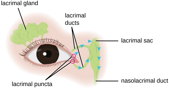
Lacrimal Apparatus

  • Lacrimal gland:
    • Serous; releases aqueous layer of tear fluid directly onto the eyeball
    • Minimizes friction, protects and cleans the eye (“tear film”)
    • Located on upper lateral aspect of orbit, drains via lacrimal ducts to the superior conjunctival fornix Fornix Vagina, Vulva, and Pelvic Floor: Anatomy
    • Innervated by parasympathetic fibers of the facial nerve Facial nerve The 7th cranial nerve. The facial nerve has two parts, the larger motor root which may be called the facial nerve proper, and the smaller intermediate or sensory root. Together they provide efferent innervation to the muscles of facial expression and to the lacrimal and salivary glands, and convey afferent information for taste from the anterior two-thirds of the tongue and for touch from the external ear. The 12 Cranial Nerves: Overview and Functions (VII) via the pterygopalatine ganglion
  • Tears:
    • Isotonic Isotonic Solutions having the same osmotic pressure as blood serum, or another solution with which they are compared. Renal Sodium and Water Regulation solution
    • Contains bactericidal Bactericidal Penicillins enzymes Enzymes Enzymes are complex protein biocatalysts that accelerate chemical reactions without being consumed by them. Due to the body’s constant metabolic needs, the absence of enzymes would make life unsustainable, as reactions would occur too slowly without these molecules. Basics of Enzymes (lysozyme and lactoferrin), immunoglobulin A, and lipocalin
  • Tarsal or Meibomian glands:
    • Sebaceous glands; produce meibum Meibum Blepharitis, which decreases evaporation of the tear film
    • Located within tarsal plate of eyelids Eyelids Each of the upper and lower folds of skin which cover the eye when closed. Blepharitis (within the superior eyelid) with orifices at the rim of the marginal zone Marginal zone MALT Lymphoma of the conjunctiva Conjunctiva The mucous membrane that covers the posterior surface of the eyelids and the anterior pericorneal surface of the eyeball. Eye: Anatomy
  • Tear drainage system Tear Drainage System Dacryocystitis—consists of:
Orbit lacrimal apparatus

Schematic diagram of the location of the lacrimal gland and apparatus

Image: “Microbiology” by CNX OpenStax. License: CC BY 4.0

Clinicopathologic Correlations

Nerve palsy Palsy paralysis of an area of the body, thus incapable of voluntary movement Cranial Nerve Palsies Causes Symptoms involving affected eye
Oculomotor (CN III)
  • Infarction of midbrain Midbrain The middle of the three primitive cerebral vesicles of the embryonic brain. Without further subdivision, midbrain develops into a short, constricted portion connecting the pons and the diencephalon. Midbrain contains two major parts, the dorsal tectum mesencephali and the ventral tegmentum mesencephali, housing components of auditory, visual, and other sensorimotor systems. Brain Stem: Anatomy
  • Berry aneurysm Berry Aneurysm Subarachnoid Hemorrhage at junction of posterior communicating and internal carotid arteries Carotid Arteries Either of the two principal arteries on both sides of the neck that supply blood to the head and neck; each divides into two branches, the internal carotid artery and the external carotid artery. Carotid Arterial System: Anatomy
  • Lesions of cavernous sinuses Cavernous sinuses An irregularly shaped venous space in the dura mater at either side of the sphenoid bone. Penis: Anatomy (neoplasm, vascular, or inflammatory)
Trochlear (CN IV)
  • Head trauma Head trauma Head trauma occurs when external forces are directed to the skull and brain structures, resulting in damage to the skull, brain, and intracranial structures. Head injuries can be classified as open (penetrating) or closed (blunt), and primary (from the initial trauma) or secondary (indirect brain injury), and range from mild to severe and life-threatening. Head Trauma
  • Tumor Tumor Inflammation at base of skull Skull The skull (cranium) is the skeletal structure of the head supporting the face and forming a protective cavity for the brain. The skull consists of 22 bones divided into the viscerocranium (facial skeleton) and the neurocranium. Skull: Anatomy
  • Microvasculopathy
  • Idiopathic Idiopathic Dermatomyositis
Eye is up and in
Abducens (CN VI)
  • Postviral syndrome
  • Ischemic mononeuropathy Mononeuropathy Disease or trauma involving a single peripheral nerve in isolation, or out of proportion to evidence of diffuse peripheral nerve dysfunction. Mononeuropathy multiplex refers to a condition characterized by multiple isolated nerve injuries. Mononeuropathies may result from a wide variety of causes, including ischemia; traumatic injury; compression; connective tissue diseases; cumulative trauma disorders; and other conditions. Mononeuropathy and Plexopathy
Eye directed medially
  • Orbital and preseptal cellulitis Preseptal cellulitis Orbital and preseptal cellulitis are infections differentiated by the anatomic sites affected in the orbit. Infection anterior to the orbital septum is preseptal cellulitis. Inoculation with the pathogen can occur through trauma or surgery. Cellulitis also occurs via extension from a nearby structure (such as from sinus infection or sinusitis). Orbital and Preseptal Cellulitis: possible complications to paranasal sinusitis Sinusitis Sinusitis refers to inflammation of the mucosal lining of the paranasal sinuses. The condition usually occurs concurrently with inflammation of the nasal mucosa (rhinitis), a condition known as rhinosinusitis. Acute sinusitis is due to an upper respiratory infection caused by a viral, bacterial, or fungal agent. Sinusitis or trauma to the eye orbit. Patients Patients Individuals participating in the health care system for the purpose of receiving therapeutic, diagnostic, or preventive procedures. Clinician–Patient Relationship may present with fever Fever Fever is defined as a measured body temperature of at least 38°C (100.4°F). Fever is caused by circulating endogenous and/or exogenous pyrogens that increase levels of prostaglandin E2 in the hypothalamus. Fever is commonly associated with chills, rigors, sweating, and flushing of the skin. Fever, malaise Malaise Tick-borne Encephalitis Virus, proptosis Proptosis Retinoblastoma ophthalmoplegia Ophthalmoplegia Paralysis of one or more of the ocular muscles due to disorders of the eye muscles, neuromuscular junction, supporting soft tissue, tendons, or innervation to the muscles. Orbital and Preseptal Cellulitis, toxic shock Shock Shock is a life-threatening condition associated with impaired circulation that results in tissue hypoxia. The different types of shock are based on the underlying cause: distributive (↑ cardiac output (CO), ↓ systemic vascular resistance (SVR)), cardiogenic (↓ CO, ↑ SVR), hypovolemic (↓ CO, ↑ SVR), obstructive (↓ CO), and mixed. Types of Shock, and impaired vision Vision Ophthalmic Exam.
  • Orbital fractures Orbital fractures An orbital fracture is a break in the continuity of one or multiple bones of the eye socket, caused by direct or indirect trauma to the orbit. Patients frequently present with lacerations around the eye, orbital pain, edema, ecchymosis, diplopia on upward gaze, numbness around the eye, and signs of muscle entrapment. Orbital Fractures: fractures to the eye sockets are classified as orbital rim Orbital Rim Orbital Fractures fractures, direct orbital floor fracture Orbital Floor Fracture Orbital Fractures, and blowout fractures. These fractures can present with blurry, decreased, or double vision Vision Ophthalmic Exam, and black-and-blue bruising around the eyes.
  • Dacryoadenitis: an inflammation Inflammation Inflammation is a complex set of responses to infection and injury involving leukocytes as the principal cellular mediators in the body’s defense against pathogenic organisms. Inflammation is also seen as a response to tissue injury in the process of wound healing. The 5 cardinal signs of inflammation are pain, heat, redness, swelling, and loss of function. Inflammation of the lacrimal glands Lacrimal Glands Dacryocystitis commonly due to a bacterial or viral infection. May present as swelling Swelling Inflammation of the outer portion of the upper lid, with redness Redness Inflammation and tenderness, pain Pain An unpleasant sensation induced by noxious stimuli which are detected by nerve endings of nociceptive neurons. Pain: Types and Pathways in the area of swelling Swelling Inflammation, excess tearing, and swelling Swelling Inflammation of the preauricular lymph nodes Lymph Nodes They are oval or bean shaped bodies (1 – 30 mm in diameter) located along the lymphatic system. Lymphatic Drainage System: Anatomy.
  • Sjögren’s syndrome: an autoimmune disease that affects the moisture-producing glands of the body. Primary symptoms are dry mouth and dry eyes. 
  • Strabismus Strabismus Strabismus is the misalignment of the eyes while fixating the gaze on an object. Strabismus can be idiopathic, but it may also be caused by cerebral palsy, uncorrected refractive errors, and extraocular muscle or cranial nerve dysfunction. Strabismus: a condition characterized by a misalignment of the eyes. If left untreated throughout childhood, strabismus Strabismus Strabismus is the misalignment of the eyes while fixating the gaze on an object. Strabismus can be idiopathic, but it may also be caused by cerebral palsy, uncorrected refractive errors, and extraocular muscle or cranial nerve dysfunction. Strabismus may result in amblyopia Amblyopia A nonspecific term referring to impaired vision. Major subcategories include stimulus deprivation-induced amblyopia and toxic amblyopia. Stimulus deprivation-induced amblyopia is a developmental disorder of the visual cortex. A discrepancy between visual information received by the visual cortex from each eye results in abnormal cortical development. Strabismus and refractive errors may cause this condition. Toxic amblyopia is a disorder of the optic nerve which is associated with alcoholism, tobacco smoking, and other toxins and as an adverse effect of the use of some medications. Strabismus or loss of depth perception Depth perception Perception of three-dimensionality. Albinism
  • Blepharitis Blepharitis Blepharitis is an ocular condition characterized by eyelid inflammation. Anterior blepharitis involves the eyelid skin and eyelashes, while the posterior type affects the meibomian glands. Often, these conditions overlap. Blepharitis: one of the most common ocular conditions; characterized by inflammation Inflammation Inflammation is a complex set of responses to infection and injury involving leukocytes as the principal cellular mediators in the body’s defense against pathogenic organisms. Inflammation is also seen as a response to tissue injury in the process of wound healing. The 5 cardinal signs of inflammation are pain, heat, redness, swelling, and loss of function. Inflammation, scaling, reddening, and crusting of the eyelid. May also present with a burning, itching, or grainy sensation

References

  1. Bowling, B. (2020). Kanski’s clinical ophthalmology: A systematic approach (9th ed.). Elsevier.
  2. Drake, R. L., Vogl, A. W., & Mitchell, A. W. M. (2019). Gray’s anatomy for students (4th ed.). Elsevier.
  3. Dutton, J. J. (2023). Atlas of clinical and surgical orbital anatomy (3rd ed.). Elsevier.
  4. Levin, L. A., Nilsson, S. F. E., Ver Hoeve, J., & Wu, S. (2021). Adler’s physiology of the eye (12th ed.). Elsevier.
  5. Netter, F. H. (2018). Atlas of human anatomy (7th ed.). Elsevier.
  6. Riordan-Eva, P., & Augsburger, J. J. (2017). Vaughan & Asbury’s general ophthalmology (19th ed.). McGraw-Hill Education.
  7. Sadda, S. R., Sarraf, D., & Freund, K. B. (2023). Retina (6th ed.). Elsevier.
  8. Wineski, L. E. (2018). Snell’s clinical anatomy by regions (10th ed.). Wolters Kluwer.

Create your free account or log in to continue reading!

Sign up now and get free access to Lecturio with concept pages, medical videos, and questions for your medical education.

User Reviews

Unwrap New Skills This Holiday 🎄 Save 30% on all plans now!

Details