Advertisement

Advertisement

Advertisement

Advertisement

Hemophilia

The hemophilias are a group of inherited, or sometimes acquired, disorders of secondary hemostasis Secondary hemostasis The coagulation cascade is a series of reactions that ultimately generates a strong, cross-linked fibrin clot. Hemostasis due to deficiency of specific clotting factors. Hemophilia A is a deficiency of factor VIII Factor VIII Factor VIII of blood coagulation. Antihemophilic factor that is part of the factor viii/von Willebrand factor complex. Factor VIII is produced in the liver and acts in the intrinsic pathway of blood coagulation. It serves as a cofactor in factor X activation and this action is markedly enhanced by small amounts of thrombin. Hemostasis, hemophilia B a deficiency of factor IX Factor IX Storage-stable blood coagulation factor acting in the intrinsic pathway of blood coagulation. Its activated form, ixa, forms a complex with factor VIII and calcium on platelet factor 3 to activate factor X to Xa. Hemostasis, and hemophilia C a deficiency of factor XI. Patients Patients Individuals participating in the health care system for the purpose of receiving therapeutic, diagnostic, or preventive procedures. Clinician–Patient Relationship present with bleeding events that may be spontaneous or associated with minor or major trauma. Management is focused mainly on treatment of acute bleeding events and prevention of bleeding events via replacement of deficient factors.

Last updated: May 17, 2024

Editorial responsibility: Stanley Oiseth, Lindsay Jones, Evelin Maza

Advertisement

Advertisement

Advertisement

Advertisement

Advertisement

Advertisement

Overview

Definition

Hemophilias are a group of disorders of secondary hemostasis Secondary hemostasis The coagulation cascade is a series of reactions that ultimately generates a strong, cross-linked fibrin clot. Hemostasis due to the deficiency of specific clotting factors.

Epidemiology

About 400 children per year are born with hemophilia in the United States.

3 different types have been described:

  • Hemophilia A:
  • Hemophilia B:
  • Hemophilia C:
    • Affects males and females equally
    • May be autosomal recessive Autosomal recessive Autosomal inheritance, both dominant and recessive, refers to the transmission of genes from the 22 autosomal chromosomes. Autosomal recessive diseases are only expressed when 2 copies of the recessive allele are inherited. Autosomal Recessive and Autosomal Dominant Inheritance or autosomal dominant Autosomal dominant Autosomal inheritance, both dominant and recessive, refers to the transmission of genes from the 22 autosomal chromosomes. Autosomal dominant diseases are expressed when only 1 copy of the dominant allele is inherited. Autosomal Recessive and Autosomal Dominant Inheritance depending on specific mutation Mutation Genetic mutations are errors in DNA that can cause protein misfolding and dysfunction. There are various types of mutations, including chromosomal, point, frameshift, and expansion mutations. Types of Mutations
    • 1 in 1 million males and females

Etiology

Genetic mutation Mutation Genetic mutations are errors in DNA that can cause protein misfolding and dysfunction. There are various types of mutations, including chromosomal, point, frameshift, and expansion mutations. Types of Mutations resulting in deficiency of clotting factors:

  • Hemophilia A:
    • Factor VIII Factor VIII Factor VIII of blood coagulation. Antihemophilic factor that is part of the factor viii/von Willebrand factor complex. Factor VIII is produced in the liver and acts in the intrinsic pathway of blood coagulation. It serves as a cofactor in factor X activation and this action is markedly enhanced by small amounts of thrombin. Hemostasis: X-linked recessive X-Linked Recessive Duchenne Muscular Dystrophy mutations in the F8 gene Gene A category of nucleic acid sequences that function as units of heredity and which code for the basic instructions for the development, reproduction, and maintenance of organisms. Basic Terms of Genetics
    • Located on the X chromosome X chromosome The female sex chromosome, being the differential sex chromosome carried by half the male gametes and all female gametes in human and other male-heterogametic species. Basic Terms of Genetics
  • Hemophilia B (Christmas disease):
    • Factor IX Factor IX Storage-stable blood coagulation factor acting in the intrinsic pathway of blood coagulation. Its activated form, ixa, forms a complex with factor VIII and calcium on platelet factor 3 to activate factor X to Xa. Hemostasis: X-linked recessive X-Linked Recessive Duchenne Muscular Dystrophy mutations in the F9 gene Gene A category of nucleic acid sequences that function as units of heredity and which code for the basic instructions for the development, reproduction, and maintenance of organisms. Basic Terms of Genetics
    • Located on the X chromosome X chromosome The female sex chromosome, being the differential sex chromosome carried by half the male gametes and all female gametes in human and other male-heterogametic species. Basic Terms of Genetics
  • Hemophilia C:
    • Factor XI: mutations in the F11 gene Gene A category of nucleic acid sequences that function as units of heredity and which code for the basic instructions for the development, reproduction, and maintenance of organisms. Basic Terms of Genetics
    • Located on chromosome Chromosome In a prokaryotic cell or in the nucleus of a eukaryotic cell, a structure consisting of or containing DNA which carries the genetic information essential to the cell. Basic Terms of Genetics 4

Acquired hemophilia:

  • The patient has developed immune-mediated autoantibodies Autoantibodies Antibodies that react with self-antigens (autoantigens) of the organism that produced them. Blotting Techniques against clotting proteins Proteins Linear polypeptides that are synthesized on ribosomes and may be further modified, crosslinked, cleaved, or assembled into complex proteins with several subunits. The specific sequence of amino acids determines the shape the polypeptide will take, during protein folding, and the function of the protein. Energy Homeostasis (most commonly factor VIII Factor VIII Factor VIII of blood coagulation. Antihemophilic factor that is part of the factor viii/von Willebrand factor complex. Factor VIII is produced in the liver and acts in the intrinsic pathway of blood coagulation. It serves as a cofactor in factor X activation and this action is markedly enhanced by small amounts of thrombin. Hemostasis).
  • Idiopathic Idiopathic Dermatomyositis in 50%
  • Can be secondary to autoimmune disorders such as lupus, rheumatoid arthritis Arthritis Acute or chronic inflammation of joints. Osteoarthritis, multiple sclerosis Sclerosis A pathological process consisting of hardening or fibrosis of an anatomical structure, often a vessel or a nerve. Wilms Tumor, Sjögren’s syndrome, and inflammatory bowel disease 
  • Other predisposing conditions include major trauma and pregnancy Pregnancy The status during which female mammals carry their developing young (embryos or fetuses) in utero before birth, beginning from fertilization to birth. Pregnancy: Diagnosis, Physiology, and Care.

Pathophysiology

Genetics Genetics Genetics is the study of genes and their functions and behaviors. Basic Terms of Genetics

  • Mutations of F8 gene Gene A category of nucleic acid sequences that function as units of heredity and which code for the basic instructions for the development, reproduction, and maintenance of organisms. Basic Terms of Genetics on X chromosome X chromosome The female sex chromosome, being the differential sex chromosome carried by half the male gametes and all female gametes in human and other male-heterogametic species. Basic Terms of Genetics:
    • 40%–45% of severe cases of hemophilia A are caused by a major inversion of the intron 22 section on the long arm Arm The arm, or “upper arm” in common usage, is the region of the upper limb that extends from the shoulder to the elbow joint and connects inferiorly to the forearm through the cubital fossa. It is divided into 2 fascial compartments (anterior and posterior). Arm: Anatomy of the X chromosome X chromosome The female sex chromosome, being the differential sex chromosome carried by half the male gametes and all female gametes in human and other male-heterogametic species. Basic Terms of Genetics.
    • 1%–5% of severe cases of hemophilia A are caused by a major inversion of the intron 1 section on the long arm Arm The arm, or “upper arm” in common usage, is the region of the upper limb that extends from the shoulder to the elbow joint and connects inferiorly to the forearm through the cubital fossa. It is divided into 2 fascial compartments (anterior and posterior). Arm: Anatomy of the X chromosome X chromosome The female sex chromosome, being the differential sex chromosome carried by half the male gametes and all female gametes in human and other male-heterogametic species. Basic Terms of Genetics.
    • Point mutations and small deletions/insertions are present in approximately 60% of cases of hemophilia A, but no specific mutation Mutation Genetic mutations are errors in DNA that can cause protein misfolding and dysfunction. There are various types of mutations, including chromosomal, point, frameshift, and expansion mutations. Types of Mutations or deletion predominates.
  • Mutations of F9 gene Gene A category of nucleic acid sequences that function as units of heredity and which code for the basic instructions for the development, reproduction, and maintenance of organisms. Basic Terms of Genetics on X chromosome X chromosome The female sex chromosome, being the differential sex chromosome carried by half the male gametes and all female gametes in human and other male-heterogametic species. Basic Terms of Genetics:
    • Highly heterogeneous: include deletions, duplications, insertions, splice-site variants, missense variants, and nonsense variants
    • 40%–45% of severe cases of hemophilia B are caused by missense variants.
  • Mutations of F11 gene Gene A category of nucleic acid sequences that function as units of heredity and which code for the basic instructions for the development, reproduction, and maintenance of organisms. Basic Terms of Genetics on chromosome Chromosome In a prokaryotic cell or in the nucleus of a eukaryotic cell, a structure consisting of or containing DNA which carries the genetic information essential to the cell. Basic Terms of Genetics 4:
    • > 200 distinct pathogenic variants in the F11 gene Gene A category of nucleic acid sequences that function as units of heredity and which code for the basic instructions for the development, reproduction, and maintenance of organisms. Basic Terms of Genetics have been reported.
    • Phe283Leu missense mutation Missense mutation A mutation in which a codon is mutated to one directing the incorporation of a different amino acid. This substitution may result in an inactive or unstable product. Types of Mutations in exon 9 is responsible for a higher prevalence Prevalence The total number of cases of a given disease in a specified population at a designated time. It is differentiated from incidence, which refers to the number of new cases in the population at a given time. Measures of Disease Frequency among those of Ashkenazi Jewish ancestry.
F8 gene location

Location of F8 gene for coagulation factor VIII: Xq28, resulting in hemophilia A

Image: “F8 gene location” by Genome Decoration Page/NCBI. License: Public Domain
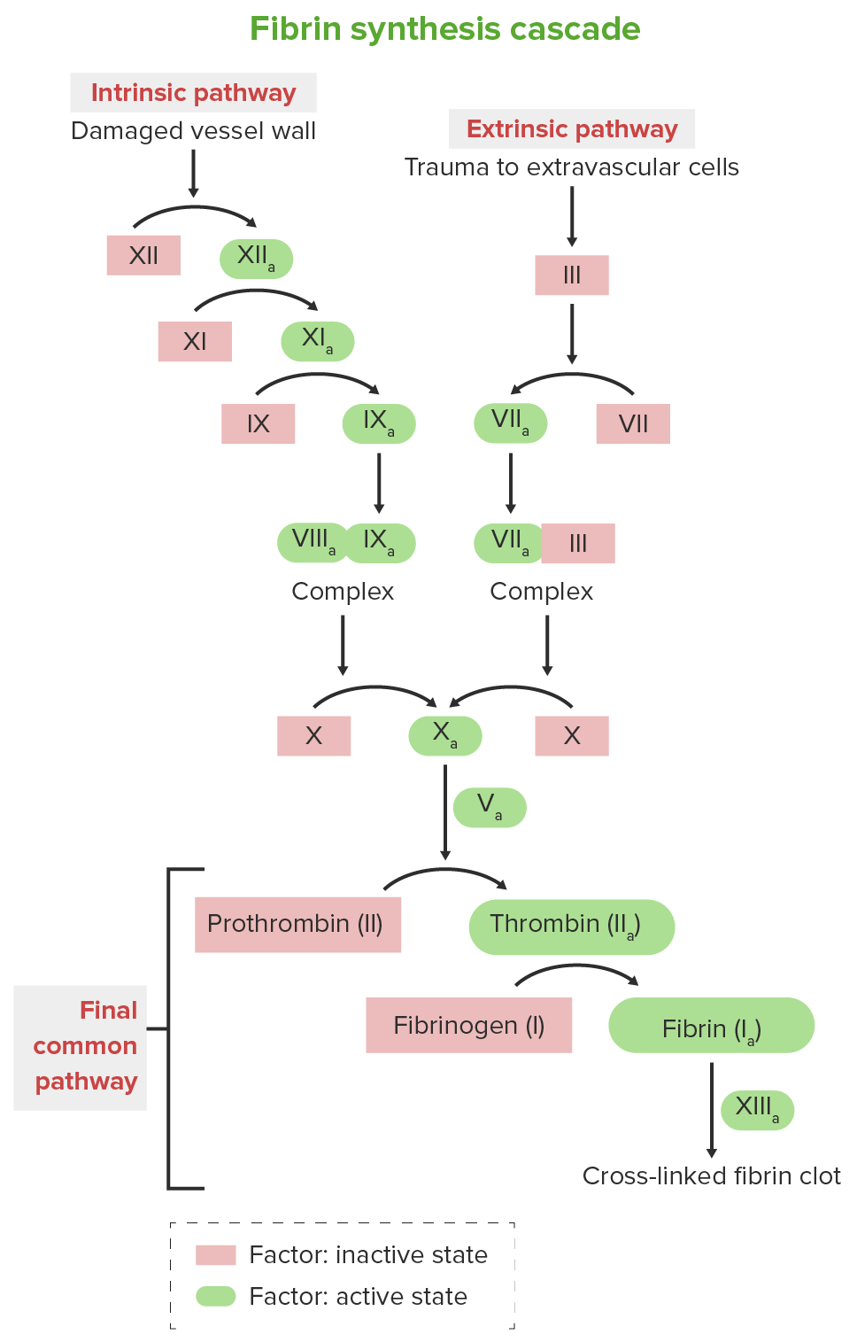
Clotting cascade

  • Produces stable fibrin Fibrin A protein derived from fibrinogen in the presence of thrombin, which forms part of the blood clot. Rapidly Progressive Glomerulonephritis clot at the site of injury
  • Two clotting pathways exist: intrinsic and extrinsic
  • The two pathways converge to a common pathway Common pathway Hemostasis; the end result is formation of a fibrin Fibrin A protein derived from fibrinogen in the presence of thrombin, which forms part of the blood clot. Rapidly Progressive Glomerulonephritis clot.
  • Factors Ⅷ, Ⅸ, and XI are components of the intrinsic pathway Intrinsic pathway The intrinsic pathway is mainly responsible for the amplification of factor X activation Hemostasis.
  • Factor deficiencies result in the dysfunction of hemostasis Hemostasis Hemostasis refers to the innate, stepwise body processes that occur following vessel injury, resulting in clot formation and cessation of bleeding. Hemostasis occurs in 2 phases, namely, primary and secondary. Primary hemostasis involves forming a plug that stops the bleeding temporarily. Secondary hemostasis involves the activation of the coagulation cascade. Hemostasis:
    • Reduced activation of prothrombin Prothrombin A plasma protein that is the inactive precursor of thrombin. It is converted to thrombin by a prothrombin activator complex consisting of factor Xa, factor V, phospholipid, and calcium ions. Hemostasis to thrombin Thrombin An enzyme formed from prothrombin that converts fibrinogen to fibrin. Hemostasis → reduced levels of thrombin Thrombin An enzyme formed from prothrombin that converts fibrinogen to fibrin. Hemostasis
    • Decreased fibrin Fibrin A protein derived from fibrinogen in the presence of thrombin, which forms part of the blood clot. Rapidly Progressive Glomerulonephritis clot formation →  delayed hemostasis Hemostasis Hemostasis refers to the innate, stepwise body processes that occur following vessel injury, resulting in clot formation and cessation of bleeding. Hemostasis occurs in 2 phases, namely, primary and secondary. Primary hemostasis involves forming a plug that stops the bleeding temporarily. Secondary hemostasis involves the activation of the coagulation cascade. Hemostasis
  • Severity of disease is proportional to severity of deficiency:
    • Mild: > 5% of normal levels
    • Moderate: 1%–5% of normal levels
    • Severe: < 1% of normal levels
Clotting cascade

Clotting cascade

Image by Lecturio.

Clinical Presentation

Manifestations

Presentation is based mainly on the severity of the factor deficiency.

Mild to moderate deficiency:

  • Presents after trauma or surgery
  • Average age at onset > 10 months, when the child begins to crawl or walk
  • Bleeding out of proportion to expected tissue response given level of trauma
  • Hemarthrosis (bleeding into joints):
    • Joint effusion Joint Effusion Septic Arthritis may lead to pain Pain An unpleasant sensation induced by noxious stimuli which are detected by nerve endings of nociceptive neurons. Pain: Types and Pathways and ↓ range of motion Range of motion The distance and direction to which a bone joint can be extended. Range of motion is a function of the condition of the joints, muscles, and connective tissues involved. Joint flexibility can be improved through appropriate muscle strength exercises. Examination of the Upper Limbs.
    • Repetitive effusions may cause early joint destruction.
  • Hematoma Hematoma A collection of blood outside the blood vessels. Hematoma can be localized in an organ, space, or tissue. Intussusception (bleeding into muscle):
    • With or without excessive bruising
    • May lead to compartment syndrome Compartment Syndrome Compartment syndrome is a surgical emergency usually occurring secondary to trauma. The condition is marked by increased pressure within a compartment that compromises the circulation and function of the tissues within that space. Compartment Syndrome

More severe deficiency:

  • Presents first in early infancy:
  • May present with spontaneous bleeding at any age
  • Nosebleeds Nosebleeds Bleeding from the nose. Granulomatosis with Polyangiitis and bleeding gums after brushing teeth Teeth Normally, an adult has 32 teeth: 16 maxillary and 16 mandibular. These teeth are divided into 4 quadrants with 8 teeth each. Each quadrant consists of 2 incisors (dentes incisivi), 1 canine (dens caninus), 2 premolars (dentes premolares), and 3 molars (dentes molares). Teeth are composed of enamel, dentin, and dental cement. Teeth: Anatomy
  • CNS (intracranial bleeding):
    • May present with altered mental status Altered Mental Status Sepsis in Children, lethargy Lethargy A general state of sluggishness, listless, or uninterested, with being tired, and having difficulty concentrating and doing simple tasks. It may be related to depression or drug addiction. Hyponatremia, vomiting Vomiting The forcible expulsion of the contents of the stomach through the mouth. Hypokalemia
    • Warrants immediate medical attention Attention Focusing on certain aspects of current experience to the exclusion of others. It is the act of heeding or taking notice or concentrating. Psychiatric Assessment 
  • GI  (bleeding into the GI tract):
    • May present with hematemesis Hematemesis Vomiting of blood that is either fresh bright red, or older ‘coffee-ground’ in character. It generally indicates bleeding of the upper gastrointestinal tract. Mallory-Weiss Syndrome (Mallory-Weiss Tear), bloody stools if gross bleeding
    • May present with symptoms/signs of anemia Anemia Anemia is a condition in which individuals have low Hb levels, which can arise from various causes. Anemia is accompanied by a reduced number of RBCs and may manifest with fatigue, shortness of breath, pallor, and weakness. Subtypes are classified by the size of RBCs, chronicity, and etiology. Anemia: Overview and Types, black stools if occult bleeding
  • Genitourinary (bleeding into the urinary tract Urinary tract The urinary tract is located in the abdomen and pelvis and consists of the kidneys, ureters, urinary bladder, and urethra. The structures permit the excretion of urine from the body. Urine flows from the kidneys through the ureters to the urinary bladder and out through the urethra. Urinary Tract: Anatomy):
    • May present with hematuria Hematuria Presence of blood in the urine. Renal Cell Carcinoma if gross bleeding
    • May present with symptoms/signs of anemia Anemia Anemia is a condition in which individuals have low Hb levels, which can arise from various causes. Anemia is accompanied by a reduced number of RBCs and may manifest with fatigue, shortness of breath, pallor, and weakness. Subtypes are classified by the size of RBCs, chronicity, and etiology. Anemia: Overview and Types if occult bleeding

Long-term complications

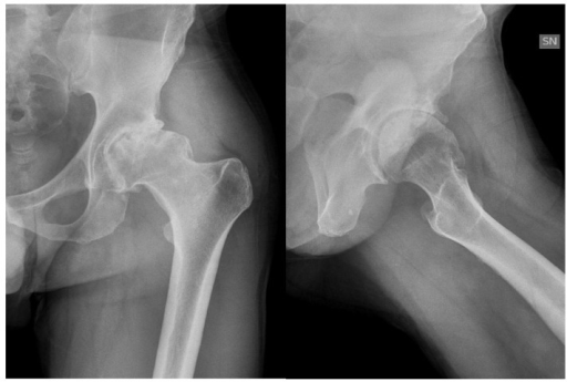
  • Hemophilic arthropathy Arthropathy Osteoarthritis:
    • Components of blood are caustic to the synovium.
    • Causes progressive joint destruction, loss of range of motion Range of motion The distance and direction to which a bone joint can be extended. Range of motion is a function of the condition of the joints, muscles, and connective tissues involved. Joint flexibility can be improved through appropriate muscle strength exercises. Examination of the Upper Limbs
  • Infections Infections Invasion of the host organism by microorganisms or their toxins or by parasites that can cause pathological conditions or diseases. Chronic Granulomatous Disease associated with transfusion of blood products:
    • Rare with appropriate screening Screening Preoperative Care precautions, which were instituted in 1985
    • Many surviving adults who received transfusions prior to that are infected with bloodborne pathogens
  • Development of factor VIII Factor VIII Factor VIII of blood coagulation. Antihemophilic factor that is part of the factor viii/von Willebrand factor complex. Factor VIII is produced in the liver and acts in the intrinsic pathway of blood coagulation. It serves as a cofactor in factor X activation and this action is markedly enhanced by small amounts of thrombin. Hemostasis autoantibodies Autoantibodies Antibodies that react with self-antigens (autoantigens) of the organism that produced them. Blotting Techniques ( factor VIII Factor VIII Factor VIII of blood coagulation. Antihemophilic factor that is part of the factor viii/von Willebrand factor complex. Factor VIII is produced in the liver and acts in the intrinsic pathway of blood coagulation. It serves as a cofactor in factor X activation and this action is markedly enhanced by small amounts of thrombin. Hemostasis inhibitors) after repetitive transfusions:
    • Patients Patients Individuals participating in the health care system for the purpose of receiving therapeutic, diagnostic, or preventive procedures. Clinician–Patient Relationship develop an “ allergy Allergy An abnormal adaptive immune response that may or may not involve antigen-specific IgE Type I Hypersensitivity Reaction” to potentially lifesaving treatment → increased susceptibility to severe bleeding complications
    • Can indirectly lead to delayed growth, delayed sexual development

Diagnosis

The diagnosis of hemophilia is made on the basis of clinical suspicion (usually due to a bleeding event) followed by demonstration of deficient factor levels (< 40%).

History

Physical examination

  • Ecchymoses
  • Hemarthroses
  • Hematomas 

Laboratory evaluation

  • CBC:
    • Normal to decreased hemoglobin/ hematocrit Hematocrit The volume of packed red blood cells in a blood specimen. The volume is measured by centrifugation in a tube with graduated markings, or with automated blood cell counters. It is an indicator of erythrocyte status in disease. For example, anemia shows a low value; polycythemia, a high value. Neonatal Polycythemia depending on occult or gross bleeding
    • Normal platelet count 
  • Coagulation studies Coagulation studies Coagulation studies are a group of hematologic laboratory studies that reflect the function of blood vessels, platelets, and coagulation factors, which all interact with one another to achieve hemostasis. Coagulation studies are usually ordered to evaluate patients with bleeding or hypercoagulation disorders. Coagulation Studies:
    • Normal bleeding time Bleeding time Duration of blood flow after skin puncture. This test is used as a measure of capillary and platelet function. Hemostasis
    • Normal PT 
    • Elevated aPTT: intrinsic pathway Intrinsic pathway The intrinsic pathway is mainly responsible for the amplification of factor X activation Hemostasis
    • Low serum levels of factor VIII Factor VIII Factor VIII of blood coagulation. Antihemophilic factor that is part of the factor viii/von Willebrand factor complex. Factor VIII is produced in the liver and acts in the intrinsic pathway of blood coagulation. It serves as a cofactor in factor X activation and this action is markedly enhanced by small amounts of thrombin. Hemostasis, IX, or XI (factor will be < 40% normal range)
    • Check von Willebrand’s factor (VWF) if factor VIII Factor VIII Factor VIII of blood coagulation. Antihemophilic factor that is part of the factor viii/von Willebrand factor complex. Factor VIII is produced in the liver and acts in the intrinsic pathway of blood coagulation. It serves as a cofactor in factor X activation and this action is markedly enhanced by small amounts of thrombin. Hemostasis levels are low to rule out von Willebrand’s disease
  • Genetic testing Genetic Testing Detection of a mutation; genotype; karyotype; or specific alleles associated with genetic traits, heritable diseases, or predisposition to a disease, or that may lead to the disease in descendants. It includes prenatal genetic testing. Myotonic Dystrophies:
    • Genetic testing Genetic Testing Detection of a mutation; genotype; karyotype; or specific alleles associated with genetic traits, heritable diseases, or predisposition to a disease, or that may lead to the disease in descendants. It includes prenatal genetic testing. Myotonic Dystrophies is not needed to diagnose hemophilia.
    • Generally reserved for female first-degree relatives for the purposes of family planning/ genetic counseling Genetic Counseling An educational process that provides information and advice to individuals or families about a genetic condition that may affect them. The purpose is to help individuals make informed decisions about marriage, reproduction, and other health management issues based on information about the genetic disease, the available diagnostic tests, and management programs. Psychosocial support is usually offered. Myotonic Dystrophies
  • Imaging:
    • CT of brain Brain The part of central nervous system that is contained within the skull (cranium). Arising from the neural tube, the embryonic brain is comprised of three major parts including prosencephalon (the forebrain); mesencephalon (the midbrain); and rhombencephalon (the hindbrain). The developed brain consists of cerebrum; cerebellum; and other structures in the brain stem. Nervous System: Anatomy, Structure, and Classification: evaluate for intracranial bleeding 
    • CT of abdomen: evaluate for retroperitoneal Retroperitoneal Peritoneum: Anatomy bleeding/ hematoma Hematoma A collection of blood outside the blood vessels. Hematoma can be localized in an organ, space, or tissue. Intussusception
    • Ultrasonography of abdomen: evaluate for retroperitoneal Retroperitoneal Peritoneum: Anatomy bleeding/ hematoma Hematoma A collection of blood outside the blood vessels. Hematoma can be localized in an organ, space, or tissue. Intussusception
    • Ultrasonography of joint: evaluate for joint swelling Swelling Inflammation/hemarthrosis

Management

General considerations

  • Genetic counseling Genetic Counseling An educational process that provides information and advice to individuals or families about a genetic condition that may affect them. The purpose is to help individuals make informed decisions about marriage, reproduction, and other health management issues based on information about the genetic disease, the available diagnostic tests, and management programs. Psychosocial support is usually offered. Myotonic Dystrophies
  • Careful surgical and/or obstetric planning
  • Avoid contact sports.
  • Avoid drugs that hamper platelet function ( aspirin Aspirin The prototypical analgesic used in the treatment of mild to moderate pain. It has anti-inflammatory and antipyretic properties and acts as an inhibitor of cyclooxygenase which results in the inhibition of the biosynthesis of prostaglandins. Aspirin also inhibits platelet aggregation and is used in the prevention of arterial and venous thrombosis. Nonsteroidal Antiinflammatory Drugs (NSAIDs), NSAIDs NSAIDS Primary vs Secondary Headaches).
  • Pain Pain An unpleasant sensation induced by noxious stimuli which are detected by nerve endings of nociceptive neurons. Pain: Types and Pathways management in selected cases

Chronic prophylactic deficiency correction for patients Patients Individuals participating in the health care system for the purpose of receiving therapeutic, diagnostic, or preventive procedures. Clinician–Patient Relationship with severe deficiency

  • Chronic infusions of deficient factor to maintain levels above the critical range:
    • 2–3 transfusions/week (though longer-acting formulations are available)
    • Goal is to maintain levels at > 1%.
    • Recombinant factor VIII Factor VIII Factor VIII of blood coagulation. Antihemophilic factor that is part of the factor viii/von Willebrand factor complex. Factor VIII is produced in the liver and acts in the intrinsic pathway of blood coagulation. It serves as a cofactor in factor X activation and this action is markedly enhanced by small amounts of thrombin. Hemostasis concentrate for hemophilia A
    • Recombinant factor IX Factor IX Storage-stable blood coagulation factor acting in the intrinsic pathway of blood coagulation. Its activated form, ixa, forms a complex with factor VIII and calcium on platelet factor 3 to activate factor X to Xa. Hemostasis concentrate for hemophilia B
    • Factor XI concentrate or fresh frozen plasma Fresh Frozen Plasma Transfusion Products ( FFP FFP Transfusion Products) for hemophilia C 
  • Desmopressin for mild hemophilia A: Desmopressin increases endothelial release of factor VIII Factor VIII Factor VIII of blood coagulation. Antihemophilic factor that is part of the factor viii/von Willebrand factor complex. Factor VIII is produced in the liver and acts in the intrinsic pathway of blood coagulation. It serves as a cofactor in factor X activation and this action is markedly enhanced by small amounts of thrombin. Hemostasis, but not factor IX Factor IX Storage-stable blood coagulation factor acting in the intrinsic pathway of blood coagulation. Its activated form, ixa, forms a complex with factor VIII and calcium on platelet factor 3 to activate factor X to Xa. Hemostasis, and VWF.
  • Antifibrinolytic agents: epsilon-amino caproic acid or tranexamic acid to control local bleeding (bleeding in the gums or GI tract and during oral surgery)
  • Monoclonal antibody emicizumab approved by the Food and Drug Administration (FDA) for prophylaxis Prophylaxis Cephalosporins of bleeding in hemophilia A:
    • Not associated with the development of factor VIII Factor VIII Factor VIII of blood coagulation. Antihemophilic factor that is part of the factor viii/von Willebrand factor complex. Factor VIII is produced in the liver and acts in the intrinsic pathway of blood coagulation. It serves as a cofactor in factor X activation and this action is markedly enhanced by small amounts of thrombin. Hemostasis inhibitors
    • Limited coverage by 3rd-party payers, cost, and limited availability restrict usage.

Acute bleeding episodes

  • Immediate transfusion of the deficient factor:
    • Recombinant factor VIII Factor VIII Factor VIII of blood coagulation. Antihemophilic factor that is part of the factor viii/von Willebrand factor complex. Factor VIII is produced in the liver and acts in the intrinsic pathway of blood coagulation. It serves as a cofactor in factor X activation and this action is markedly enhanced by small amounts of thrombin. Hemostasis concentrate for hemophilia A
    • Recombinant factor IX Factor IX Storage-stable blood coagulation factor acting in the intrinsic pathway of blood coagulation. Its activated form, ixa, forms a complex with factor VIII and calcium on platelet factor 3 to activate factor X to Xa. Hemostasis concentrate for hemophilia B
    • Factor XI concentrate or FFP FFP Transfusion Products for hemophilia C
  • Co-transfusion with packed RBCs Packed RBCs Transfusion Products in the setting of hemodynamic instability

Surgical evaluation/intervention

  • Intracranial hemorrhage Intracranial hemorrhage Subarachnoid hemorrhage (SAH) is a type of cerebrovascular accident (stroke) resulting from intracranial hemorrhage into the subarachnoid space between the arachnoid and the pia mater layers of the meninges surrounding the brain. Most sahs originate from a saccular aneurysm in the circle of willis but may also occur as a result of trauma, uncontrolled hypertension, vasculitis, anticoagulant use, or stimulant use. Subarachnoid Hemorrhage 
  • Compartment syndrome Compartment Syndrome Compartment syndrome is a surgical emergency usually occurring secondary to trauma. The condition is marked by increased pressure within a compartment that compromises the circulation and function of the tissues within that space. Compartment Syndrome 
  • Hematoma Hematoma A collection of blood outside the blood vessels. Hematoma can be localized in an organ, space, or tissue. Intussusception debridement Debridement The removal of foreign material and devitalized or contaminated tissue from or adjacent to a traumatic or infected lesion until surrounding healthy tissue is exposed. Stevens-Johnson Syndrome/debulking/evacuation 
  • Arthrocentesis Arthrocentesis Puncture and aspiration of fluid (e.g., synovial fluid) from a joint cavity. It is used sometimes to irrigate or administer drugs into a joint cavity. Septic Arthritis, joint replacement

Prognosis Prognosis A prediction of the probable outcome of a disease based on a individual’s condition and the usual course of the disease as seen in similar situations. Non-Hodgkin Lymphomas

  • Severe hemophilia results in a limited life expectancy Life expectancy Based on known statistical data, the number of years which any person of a given age may reasonably expected to live. Population Pyramids.
  • Availability of factor concentrates → dramatic improvement in life expectancy Life expectancy Based on known statistical data, the number of years which any person of a given age may reasonably expected to live. Population Pyramids and in quality Quality Activities and programs intended to assure or improve the quality of care in either a defined medical setting or a program. The concept includes the assessment or evaluation of the quality of care; identification of problems or shortcomings in the delivery of care; designing activities to overcome these deficiencies; and follow-up monitoring to ensure effectiveness of corrective steps. Quality Measurement and Improvement of life 
  • Life expectancy Life expectancy Based on known statistical data, the number of years which any person of a given age may reasonably expected to live. Population Pyramids for hemophilia patients Patients Individuals participating in the health care system for the purpose of receiving therapeutic, diagnostic, or preventive procedures. Clinician–Patient Relationship born after 1985 is around 65 years.
  • The most common cause of death is liver failure Liver failure Severe inability of the liver to perform its normal metabolic functions, as evidenced by severe jaundice and abnormal serum levels of ammonia; bilirubin; alkaline phosphatase; aspartate aminotransferase; lactate dehydrogenases; and albumin/globulin ratio. Autoimmune Hepatitis.
  • Complications of HIV HIV Anti-HIV Drugs and hepatitis C Hepatitis C Hepatitis C is an infection of the liver caused by the hepatitis C virus (HCV). The infection can be transmitted through infectious blood or body fluids and may be transmitted during childbirth or through IV drug use or sexual intercourse. Hepatitis C virus can cause both acute and chronic hepatitis, ranging from a mild to a serious, lifelong illness including liver cirrhosis and hepatocellular carcinoma (HCC). Hepatitis C Virus infection remain the most important cause of death in patients Patients Individuals participating in the health care system for the purpose of receiving therapeutic, diagnostic, or preventive procedures. Clinician–Patient Relationship born prior to 1985.

Differential Diagnosis

  • Liver failure Liver failure Severe inability of the liver to perform its normal metabolic functions, as evidenced by severe jaundice and abnormal serum levels of ammonia; bilirubin; alkaline phosphatase; aspartate aminotransferase; lactate dehydrogenases; and albumin/globulin ratio. Autoimmune Hepatitis: The liver Liver The liver is the largest gland in the human body. The liver is found in the superior right quadrant of the abdomen and weighs approximately 1.5 kilograms. Its main functions are detoxification, metabolism, nutrient storage (e.g., iron and vitamins), synthesis of coagulation factors, formation of bile, filtration, and storage of blood. Liver: Anatomy is the site for synthesis Synthesis Polymerase Chain Reaction (PCR) of coagulation factors Coagulation factors Endogenous substances, usually proteins, that are involved in the blood coagulation process. Hemostasis; thus, a dysfunctional liver Liver The liver is the largest gland in the human body. The liver is found in the superior right quadrant of the abdomen and weighs approximately 1.5 kilograms. Its main functions are detoxification, metabolism, nutrient storage (e.g., iron and vitamins), synthesis of coagulation factors, formation of bile, filtration, and storage of blood. Liver: Anatomy would result in deficiency in the clotting factors. Failure would be diagnosed clinically in the setting of liver Liver The liver is the largest gland in the human body. The liver is found in the superior right quadrant of the abdomen and weighs approximately 1.5 kilograms. Its main functions are detoxification, metabolism, nutrient storage (e.g., iron and vitamins), synthesis of coagulation factors, formation of bile, filtration, and storage of blood. Liver: Anatomy disease with confirmation of the nature of the bleeding diathesis Bleeding diathesis Wiskott-Aldrich Syndrome via laboratory workup. Treatment is aimed at controlling bleeding, replacing deficient clotting factors, and addressing the underlying cause of liver Liver The liver is the largest gland in the human body. The liver is found in the superior right quadrant of the abdomen and weighs approximately 1.5 kilograms. Its main functions are detoxification, metabolism, nutrient storage (e.g., iron and vitamins), synthesis of coagulation factors, formation of bile, filtration, and storage of blood. Liver: Anatomy disease. 
  • Vitamin K deficiency Vitamin K Deficiency A nutritional condition produced by a deficiency of vitamin K in the diet, characterized by an increased tendency to hemorrhage (hemorrhagic disorders). Such bleeding episodes may be particularly severe in newborn infants. Fat-soluble Vitamins and their Deficiencies: Vitamin K Vitamin K A lipid cofactor that is required for normal blood clotting. Several forms of vitamin K have been identified: vitamin K 1 (phytomenadione) derived from plants, vitamin K 2 (menaquinone) from bacteria, and synthetic naphthoquinone provitamins, vitamin K 3 (menadione). Vitamin k 3 provitamins, after being alkylated in vivo, exhibit the antifibrinolytic activity of vitamin k. Green leafy vegetables, liver, cheese, butter, and egg yolk are good sources of vitamin k. Fat-soluble Vitamins and their Deficiencies is needed for the activation of clotting factors II, VII, IX, and X; thus, a deficiency would lead to inactivation of these factors and present with bleeding complications. Diagnosis is made by laboratory workup demonstrating low vitamin K Vitamin K A lipid cofactor that is required for normal blood clotting. Several forms of vitamin K have been identified: vitamin K 1 (phytomenadione) derived from plants, vitamin K 2 (menaquinone) from bacteria, and synthetic naphthoquinone provitamins, vitamin K 3 (menadione). Vitamin k 3 provitamins, after being alkylated in vivo, exhibit the antifibrinolytic activity of vitamin k. Green leafy vegetables, liver, cheese, butter, and egg yolk are good sources of vitamin k. Fat-soluble Vitamins and their Deficiencies levels and downstream deficiency of associated clotting factors. Treatment is aimed at control of bleeding and correct  vitamin K deficiency Vitamin K Deficiency A nutritional condition produced by a deficiency of vitamin K in the diet, characterized by an increased tendency to hemorrhage (hemorrhagic disorders). Such bleeding episodes may be particularly severe in newborn infants. Fat-soluble Vitamins and their Deficiencies and/or deficient clotting factors.
  • Disseminated intravascular coagulation Disseminated intravascular coagulation Disseminated intravascular coagulation (DIC) is a condition characterized by systemic bodywide activation of the coagulation cascade. This cascade results in both widespread microvascular thrombi contributing to multiple organ dysfunction and consumption of clotting factors and platelets, leading to hemorrhage. Disseminated Intravascular Coagulation ( DIC DIC Disseminated intravascular coagulation (DIC) is a condition characterized by systemic bodywide activation of the coagulation cascade. This cascade results in both widespread microvascular thrombi contributing to multiple organ dysfunction and consumption of clotting factors and platelets, leading to hemorrhage. Disseminated Intravascular Coagulation): Disseminated intravascular coagulation Disseminated intravascular coagulation Disseminated intravascular coagulation (DIC) is a condition characterized by systemic bodywide activation of the coagulation cascade. This cascade results in both widespread microvascular thrombi contributing to multiple organ dysfunction and consumption of clotting factors and platelets, leading to hemorrhage. Disseminated Intravascular Coagulation, an imbalance between native prothrombotic and antithrombotic factors, is a complication of many diseases. An inciting event causes widespread thrombosis Thrombosis Formation and development of a thrombus or blood clot in the blood vessel. Epidemic Typhus (exhausting available clotting factor reserves) followed by uncontrolled bleeding. It has high morbidity Morbidity The proportion of patients with a particular disease during a given year per given unit of population. Measures of Health Status and mortality Mortality All deaths reported in a given population. Measures of Health Status and thus requires critical care intervention. Treatment is aimed at control of bleeding, resuscitation Resuscitation The restoration to life or consciousness of one apparently dead. . Neonatal Respiratory Distress Syndrome to maintain hemodynamic stability, and addressing the inciting event.  
  • Thrombotic thrombocytopenic purpura Thrombotic thrombocytopenic purpura Thrombotic thrombocytopenic purpura (TTP) is a life-threatening condition due to either a congenital or an acquired deficiency of ADAMTS-13, a metalloproteinase that cleaves multimers of von Willebrand factor (VWF). The large multimers then aggregate excessive platelets resulting in microvascular thrombosis and an increase in consumption of platelets. Thrombotic Thrombocytopenic Purpura ( TTP TTP Thrombotic thrombocytopenic purpura (TTP) is a life-threatening condition due to either a congenital or an acquired deficiency of adamts-13, a metalloproteinase that cleaves multimers of von Willebrand factor (vWF). The large multimers then aggregate excessive platelets resulting in microvascular thrombosis and an increase in consumption of platelets. Thrombotic Thrombocytopenic Purpura): a disorder due to a deficiency in the metalloproteinase responsible for breakdown of VWF, which can be congenital Congenital Chorioretinitis or acquired: Thrombotic thrombocytopenic purpura Thrombotic thrombocytopenic purpura Thrombotic thrombocytopenic purpura (TTP) is a life-threatening condition due to either a congenital or an acquired deficiency of ADAMTS-13, a metalloproteinase that cleaves multimers of von Willebrand factor (VWF). The large multimers then aggregate excessive platelets resulting in microvascular thrombosis and an increase in consumption of platelets. Thrombotic Thrombocytopenic Purpura falls into the differential diagnosis with idiopathic Idiopathic Dermatomyositis thrombocytopenic purpura ( ITP ITP Immune thrombocytopenic purpura (ITP), formerly known as idiopathic thrombocytopenic purpura, is a condition that develops secondary to immune-mediated destruction of platelets, resulting in thrombocytopenia (platelet count < 100,000/mm³). Immune thrombocytopenic purpura can be either primary or secondary due to drugs or underlying disease. Immune Thrombocytopenic Purpura) and DIC DIC Disseminated intravascular coagulation (DIC) is a condition characterized by systemic bodywide activation of the coagulation cascade. This cascade results in both widespread microvascular thrombi contributing to multiple organ dysfunction and consumption of clotting factors and platelets, leading to hemorrhage. Disseminated Intravascular Coagulation, all presenting with thrombocytopenia Thrombocytopenia Thrombocytopenia occurs when the platelet count is < 150,000 per microliter. The normal range for platelets is usually 150,000-450,000/µL of whole blood. Thrombocytopenia can be a result of decreased production, increased destruction, or splenic sequestration of platelets. Patients are often asymptomatic until platelet counts are < 50,000/µL. Thrombocytopenia. Thrombotic thrombocytopenic purpura Thrombotic thrombocytopenic purpura Thrombotic thrombocytopenic purpura (TTP) is a life-threatening condition due to either a congenital or an acquired deficiency of ADAMTS-13, a metalloproteinase that cleaves multimers of von Willebrand factor (VWF). The large multimers then aggregate excessive platelets resulting in microvascular thrombosis and an increase in consumption of platelets. Thrombotic Thrombocytopenic Purpura is more acute than hemophilia and requires urgent plasmapheresis Plasmapheresis Procedure whereby plasma is separated and extracted from anticoagulated whole blood and the red cells retransfused to the donor. Plasmapheresis is also employed for therapeutic use. Stevens-Johnson Syndrome.
  • Henoch–Schönlein purpura: an autoimmune small-vessel vasculitis Small-Vessel Vasculitis Henoch-Schönlein Purpura: Henoch–Schönlein purpura typically presents as a tetrad of abdominal pain Abdominal Pain Acute Abdomen, arthralgia Arthralgia Pain in the joint. Rheumatic Fever, purpuric rash Rash Rocky Mountain Spotted Fever, and hematuria Hematuria Presence of blood in the urine. Renal Cell Carcinoma. The disorder is diagnosed clinically and managed symptomatically.
  • Von Willebrand disease Von Willebrand disease Von Willebrand disease (vWD) is a bleeding disorder characterized by a qualitative or quantitative deficiency of von Willebrand factor (vWF). Von Willebrand factor is a multimeric protein involved in the plate adhesion phase of hemostasis by forming a bridge between platelets and damaged portions of the vessel wall. Von Willebrand Disease: an inherited autosomal bleeding disorder that can present with mild to moderate bleeding at any age: Diagnosis is made by demonstration of VWF deficiency on laboratory analysis. Treatment is with control of bleeding and administration of desmopressin and antifibrinolytic agents.

References

  1. Hoots W, Shapiro A. (2020). Hemophilia A and B: Routine management including prophylaxis. Retrieved March 6, 2021, from https://www.uptodate.com/contents/hemophilia-a-and-b-routine-management-including-prophylaxis
  2. Hoots W, Shapiro A, Heiman M. (2021). Genetics of hemophilia A and B. Retrieved March 6, 2021, from https://www.uptodate.com/contents/genetics-of-hemophilia-a-and-b
  3. Bérubé C. (2021). Factor XI (eleven) deficiency. Retrieved March 6, 2021, from https://www.uptodate.com/contents/factor-xi-eleven-deficiency
  4. Kasper D, Fauci A, Hauser S, Longo DL, Jameson J, Loscalzo J. (2012). Harrison’s Principles of Internal Medicine (18th ed.). New York: McGraw Hill Education, pp. 2180–2186.
  5. Duga S, Salomon O. (2013). Congenital factor XI deficiency: An update. Seminars in Thrombosis Hemostasis 39(6):621–631.

Create your free account or log in to continue reading!

Sign up now and get free access to Lecturio with concept pages, medical videos, and questions for your medical education.

User Reviews

Unwrap New Skills This Holiday 🎄 Save 30% on all plans now!

Details