El herpes zóster (también conocido como culebrilla) es una infección viral de reactivación causada por el virusVirusViruses are infectious, obligate intracellular parasites composed of a nucleic acid core surrounded by a protein capsid. Viruses can be either naked (non-enveloped) or enveloped. The classification of viruses is complex and based on many factors, including type and structure of the nucleoid and capsid, the presence of an envelope, the replication cycle, and the host range. Virology de la varicela-zóster. El virusVirusViruses are infectious, obligate intracellular parasites composed of a nucleic acid core surrounded by a protein capsid. Viruses can be either naked (non-enveloped) or enveloped. The classification of viruses is complex and based on many factors, including type and structure of the nucleoid and capsid, the presence of an envelope, the replication cycle, and the host range. Virology de la varicela-zóster permanece latente enENErythema nodosum is an immune-mediated panniculitis (inflammation of the subcutaneous fat) caused by a type IV (delayed-type) hypersensitivity reaction. It commonly manifests in young women as tender, erythematous nodules on the shins.Erythema Nodosum la raíz dorsal del ganglio tras la fase de infección primaria de la varicela. La edad, el estrés o losLOSNeisseria estados inmunodeprimidos pueden desencadenar la reactivación del virusVirusViruses are infectious, obligate intracellular parasites composed of a nucleic acid core surrounded by a protein capsid. Viruses can be either naked (non-enveloped) or enveloped. The classification of viruses is complex and based on many factors, including type and structure of the nucleoid and capsid, the presence of an envelope, the replication cycle, and the host range. Virology. El herpes zóster se presenta clínicamente con una distribución enENErythema nodosum is an immune-mediated panniculitis (inflammation of the subcutaneous fat) caused by a type IV (delayed-type) hypersensitivity reaction. It commonly manifests in young women as tender, erythematous nodules on the shins.Erythema Nodosum dermatoma y enENErythema nodosum is an immune-mediated panniculitis (inflammation of the subcutaneous fat) caused by a type IV (delayed-type) hypersensitivity reaction. It commonly manifests in young women as tender, erythematous nodules on the shins.Erythema Nodosum forma de una erupción dolorosa, única y unilateral. El diagnóstico se realiza principalmente a partir de losLOSNeisseria antecedentes clínicos y la exploración física. Sin embargo, pueden realizarse pruebas de laboratorio (como la PCRPCRPolymerase chain reaction (PCR) is a technique that amplifies DNA fragments exponentially for analysis. The process is highly specific, allowing for the targeting of specific genomic sequences, even with minuscule sample amounts. The PCR cycles multiple times through 3 phases: denaturation of the template DNA, annealing of a specific primer to the individual DNA strands, and synthesis/elongation of new DNA molecules.Polymerase Chain Reaction (PCR)) si el diagnóstico no está claro. El manejo incluye terapia antiviralAntiviralAntivirals for Hepatitis B y tratamiento sintomático.
El herpes zóster (también conocido como culebrilla) es una infección viral de reactivación causada por el virusVirusViruses are infectious, obligate intracellular parasites composed of a nucleic acid core surrounded by a protein capsid. Viruses can be either naked (non-enveloped) or enveloped. The classification of viruses is complex and based on many factors, including type and structure of the nucleoid and capsid, the presence of an envelope, the replication cycle, and the host range. Virology de la varicela-zóster que se manifiesta como una erupción vesicular enENErythema nodosum is an immune-mediated panniculitis (inflammation of the subcutaneous fat) caused by a type IV (delayed-type) hypersensitivity reaction. It commonly manifests in young women as tender, erythematous nodules on the shins.Erythema Nodosum un dermatoma, unilateral y dolorosa.
Epidemiología
Incidencia anual enENErythema nodosum is an immune-mediated panniculitis (inflammation of the subcutaneous fat) caused by a type IV (delayed-type) hypersensitivity reaction. It commonly manifests in young women as tender, erythematous nodules on the shins.Erythema NodosumlosLOSNeisseria Estados Unidos: aproximadamente 1 millón de casos.
Aproximadamente el 30% de la población experimentará herpes zóster a lo largo de su vida.
Más común enENErythema nodosum is an immune-mediated panniculitis (inflammation of the subcutaneous fat) caused by a type IV (delayed-type) hypersensitivity reaction. It commonly manifests in young women as tender, erythematous nodules on the shins.Erythema Nodosum:
Edad avanzada
Inmunodeprimidos
Sexo femenino
Solo individuos previamente infectados por el virusVirusViruses are infectious, obligate intracellular parasites composed of a nucleic acid core surrounded by a protein capsid. Viruses can be either naked (non-enveloped) or enveloped. The classification of viruses is complex and based on many factors, including type and structure of the nucleoid and capsid, the presence of an envelope, the replication cycle, and the host range. Virology de la varicela-zóster (por varicela o raramente, por la vacuna contra la varicela)
Etiología
Organismo causante: virusVirusViruses are infectious, obligate intracellular parasites composed of a nucleic acid core surrounded by a protein capsid. Viruses can be either naked (non-enveloped) or enveloped. The classification of viruses is complex and based on many factors, including type and structure of the nucleoid and capsid, the presence of an envelope, the replication cycle, and the host range. Virology de la varicela-zóster
También conocido como herpesvirus humano tipo 3
VirusVirusViruses are infectious, obligate intracellular parasites composed of a nucleic acid core surrounded by a protein capsid. Viruses can be either naked (non-enveloped) or enveloped. The classification of viruses is complex and based on many factors, including type and structure of the nucleoid and capsid, the presence of an envelope, the replication cycle, and the host range. Virology de ADN de doble cadena con envoltura
Pertenece a la familia de losLOSNeisseriaHerpesviridaeHerpesviridaeA family of enveloped, linear, double-stranded DNA viruses infecting a wide variety of animals. Subfamilies, based on biological characteristics, include: alphaherpesvirinae; betaherpesvirinae; and gammaherpesvirinae.Herpes Simplex Virus 1 and 2
Transmisión:
Contacto directo con las ampollas
Gotitas respiratorias (de individuos con enfermedad diseminada)
Causa la varicela enENErythema nodosum is an immune-mediated panniculitis (inflammation of the subcutaneous fat) caused by a type IV (delayed-type) hypersensitivity reaction. It commonly manifests in young women as tender, erythematous nodules on the shins.Erythema Nodosum individuos expuestos sin inmunidad
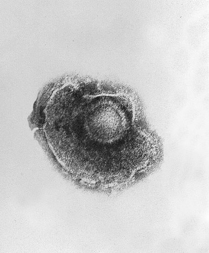
Imagen de microscopía electrónica de transmisión que muestra un único virus de la varicela-zóster (VZV), también conocido como herpesvirus humano 3, que causa la varicela
Imagen: “Ultrastructural features exhibited by a single varicella-zoster virus (VZV), also known as human herpesvirus 3 (HHV-3), the cause of chickenpox.” por CDC. Licencia: Dominio Público
Factores de riesgo
Paciente inmunodeprimido:
Trasplante
Terapia inmunosupresora
VirusVirusViruses are infectious, obligate intracellular parasites composed of a nucleic acid core surrounded by a protein capsid. Viruses can be either naked (non-enveloped) or enveloped. The classification of viruses is complex and based on many factors, including type and structure of the nucleoid and capsid, the presence of an envelope, the replication cycle, and the host range. Virology de la inmunodeficiencia humana (VIH)
Trastorno autoinmune
Enfermedad activa o crónica
Traumatismo físico
Antecedentes familiares
Edad
Estrés
Fisiopatología
El virusVirusViruses are infectious, obligate intracellular parasites composed of a nucleic acid core surrounded by a protein capsid. Viruses can be either naked (non-enveloped) or enveloped. The classification of viruses is complex and based on many factors, including type and structure of the nucleoid and capsid, the presence of an envelope, the replication cycle, and the host range. Virology de la varicela-zóster causa 2 síndromes distintos:
Infección primaria (varicela):
Se transmite a través del aerosol → se dirige a las células mucoepiteliales.
ViremiaViremiaThe presence of viruses in the blood.Erythema Infectiosum → contagiosa, se produce una enfermedad febril.
Tras la resolución, las partículas virales permanecen enENErythema nodosum is an immune-mediated panniculitis (inflammation of the subcutaneous fat) caused by a type IV (delayed-type) hypersensitivity reaction. It commonly manifests in young women as tender, erythematous nodules on the shins.Erythema NodosumlosLOSNeisseria ganglios de la raiz dorsal o enENErythema nodosum is an immune-mediated panniculitis (inflammation of the subcutaneous fat) caused by a type IV (delayed-type) hypersensitivity reaction. It commonly manifests in young women as tender, erythematous nodules on the shins.Erythema Nodosum otros ganglios sensoriales.
El sistema inmunitario del huésped suprime la replicación del virusVirusViruses are infectious, obligate intracellular parasites composed of a nucleic acid core surrounded by a protein capsid. Viruses can be either naked (non-enveloped) or enveloped. The classification of viruses is complex and based on many factors, including type and structure of the nucleoid and capsid, the presence of an envelope, the replication cycle, and the host range. Virology → permanece latente durante años o décadas (periodo de latencia).
Infección secundaria (herpes zóster):
El sistema inmunitario del huésped no logra contener el virusVirusViruses are infectious, obligate intracellular parasites composed of a nucleic acid core surrounded by a protein capsid. Viruses can be either naked (non-enveloped) or enveloped. The classification of viruses is complex and based on many factors, including type and structure of the nucleoid and capsid, the presence of an envelope, the replication cycle, and the host range. Virology → el virusVirusViruses are infectious, obligate intracellular parasites composed of a nucleic acid core surrounded by a protein capsid. Viruses can be either naked (non-enveloped) or enveloped. The classification of viruses is complex and based on many factors, including type and structure of the nucleoid and capsid, the presence of an envelope, the replication cycle, and the host range. Virology de la varicela-zóster se reactiva.
Se extiende por el nervio sensorial → piel → erupción.
Respuesta inflamatoria enENErythema nodosum is an immune-mediated panniculitis (inflammation of the subcutaneous fat) caused by a type IV (delayed-type) hypersensitivity reaction. It commonly manifests in young women as tender, erythematous nodules on the shins.Erythema NodosumlosLOSNeisseria ganglios sensoriales:
Implica a las células plasmáticas y a losLOSNeisseria linfocitos T.
Puede provocar daños neuronales → dolorDolorInflammation neuropático.
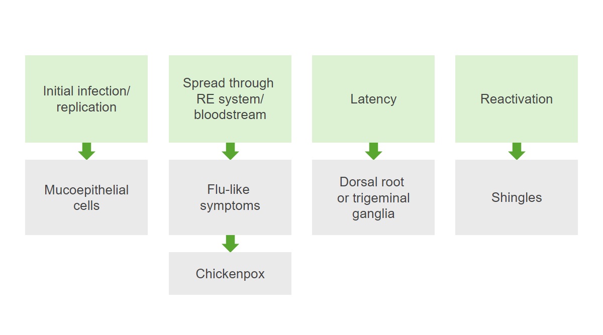
Patogénesis del virus de la varicela-zóster: La infección replica los virus en las células mucoepiteliales y luego se extiende por el sistema reticuloendotelial (RE) y el torrente sanguíneo, provocando síntomas similares a los de la gripe y la varicela. Tras la resolución de la infección primaria, se produce un periodo de latencia y el virus permanece latente en los ganglios de la raíz dorsal. La reactivación de la infección da lugar al herpes zóster.
Distribución enENErythema nodosum is an immune-mediated panniculitis (inflammation of the subcutaneous fat) caused by a type IV (delayed-type) hypersensitivity reaction. It commonly manifests in young women as tender, erythematous nodules on the shins.Erythema Nodosum dermatoma
Inicialmente aparece como pápulas eritematosas
Progresa enENErythema nodosum is an immune-mediated panniculitis (inflammation of the subcutaneous fat) caused by a type IV (delayed-type) hypersensitivity reaction. It commonly manifests in young women as tender, erythematous nodules on the shins.Erythema Nodosum bullas o grupos de vesículas
Se vuelve pustulosa o hemorrágica enENErythema nodosum is an immune-mediated panniculitis (inflammation of the subcutaneous fat) caused by a type IV (delayed-type) hypersensitivity reaction. It commonly manifests in young women as tender, erythematous nodules on the shins.Erythema Nodosum 3–4 días
Las lesiones comienzan a formar costras enENErythema nodosum is an immune-mediated panniculitis (inflammation of the subcutaneous fat) caused by a type IV (delayed-type) hypersensitivity reaction. It commonly manifests in young women as tender, erythematous nodules on the shins.Erythema Nodosum 7–10 días (ya no son infecciosas).
Pueden aparecer cicatrices e hipo o hiperpigmentación.
Síntomas sistémicos (< 20% de losLOSNeisseria casos):
Fiebre
Cefalea
Malestar
Fatiga
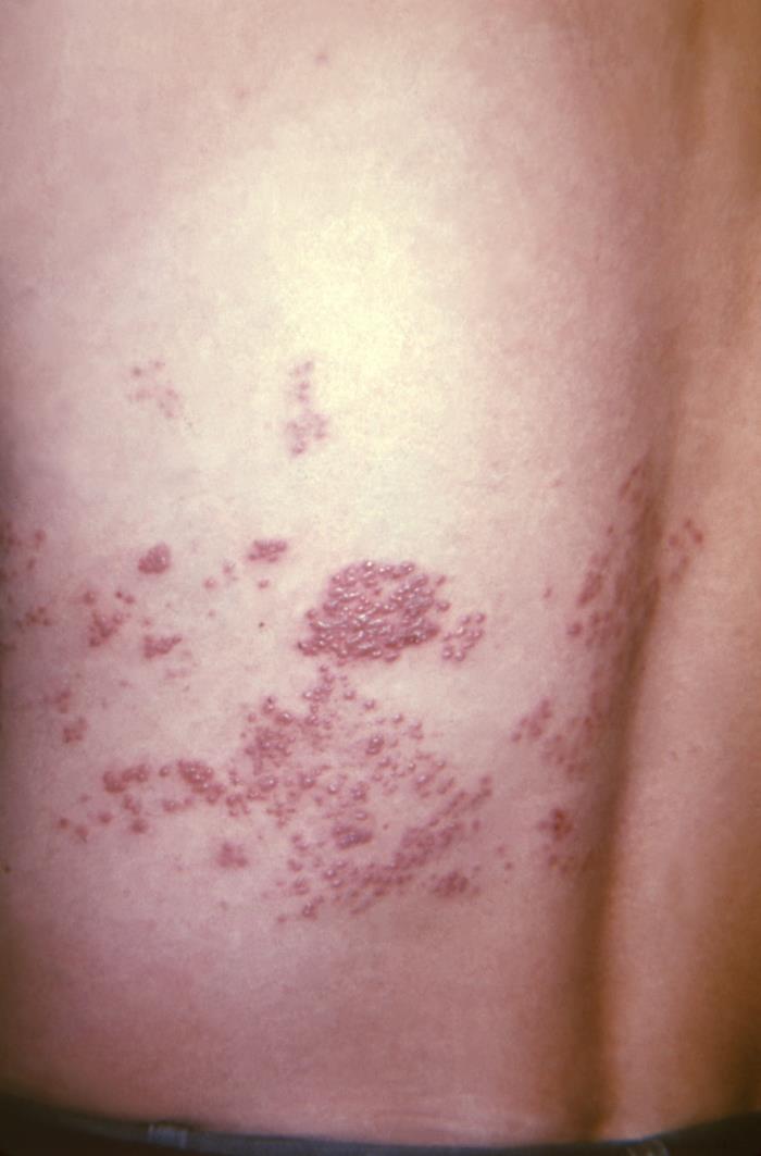
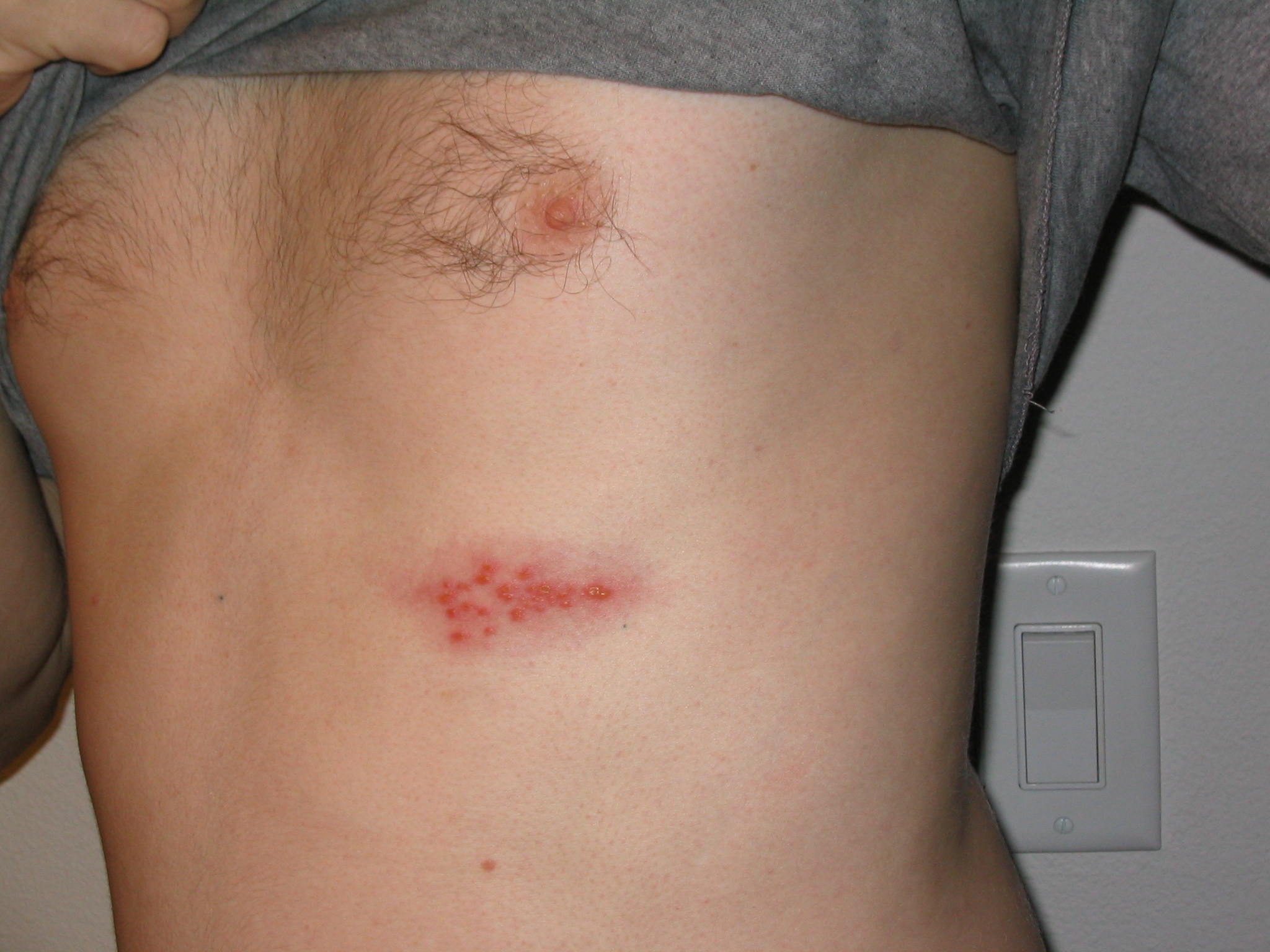
Erupción de herpes zóster en el dermatoma T10–11 a lo largo de la espalda de un paciente
Imagen: “This view of a patient’s skin, revealed a maculopapular rash, which had been due to an outbreak of shingles.” por CDC. Licencia: Dominio Público
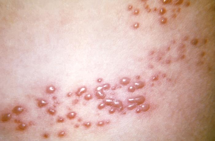
Primer plano de la erupción vesicular del herpes zóster
Imagen: “This view of a patient’s skin, revealed a maculopapular rash, which had been due to an outbreak of shingles.” por CDC. Licencia: Dominio Público
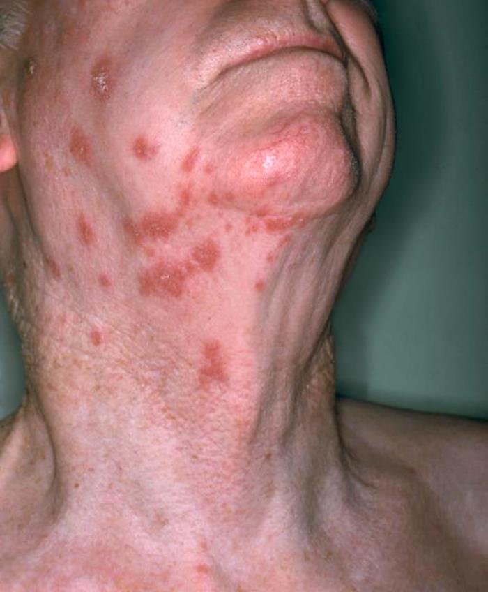
Vista anterolateral del cuello de un paciente que muestra la presencia de una erupción eritematosa debida al herpes zóster
Imagen: “Anterolateral view of this patient’s neck showing the presence of an erythematous rash due to shingles” por NIAID. Licencia: Dominio Público
Imagen de un brote de herpes en el pecho
Imagen: “Picture of a shingles (herpes zoster) outbreak on the chest” por Preston Hunt. Licencia: CC BY 3.0
Herpes zóster diseminado
Las siguientes son presentaciones graves, que pueden ocurrir enENErythema nodosum is an immune-mediated panniculitis (inflammation of the subcutaneous fat) caused by a type IV (delayed-type) hypersensitivity reaction. It commonly manifests in young women as tender, erythematous nodules on the shins.Erythema Nodosum pacientes inmunodeprimidos:
Diseminación cutánea:
Las lesiones vesiculares tienen una distribución generalizada.
Vesículas enENErythema nodosum is an immune-mediated panniculitis (inflammation of the subcutaneous fat) caused by a type IV (delayed-type) hypersensitivity reaction. It commonly manifests in young women as tender, erythematous nodules on the shins.Erythema Nodosum el canal auditivo o pabellón auricular
Características adicionales:
Disgeusia
Anomalías auditivas
Lagrimeo
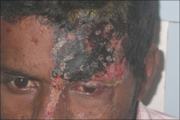
Herpes zóster oftálmico con afectación del ojo izquierdo
Imagen: “External photograph showing herpes zoster ophthalmicus” por Sudharshan S et al. Licencia: CC BY 2.0
Complicaciones
Neuralgia postherpética:
Complicación más común
Persistencia significativa del dolorDolorInflammation durante 90 días después del desarrollo de la erupción
Otros síntomas:
Adormecimiento
Disestesias
Prurito
Alodinia
NecrosisNecrosisThe death of cells in an organ or tissue due to disease, injury or failure of the blood supply.Ischemic Cell Damage retiniana aguda:
Características:
Iridociclitis aguda
Vitritis
Retinitis necrotizante
VasculitisVasculitisInflammation of any one of the blood vessels, including the arteries; veins; and rest of the vasculature system in the body.Systemic Lupus Erythematosus retiniana oclusiva
Complicaciones:
Pérdida rápida de la visión
Desprendimiento de retinaRetinaThe ten-layered nervous tissue membrane of the eye. It is continuous with the optic nerve and receives images of external objects and transmits visual impulses to the brain. Its outer surface is in contact with the choroid and the inner surface with the vitreous body. The outermost layer is pigmented, whereas the inner nine layers are transparent.Eye: Anatomy
Otras complicaciones:
Infecciones bacterianas secundarias de la piel
MeningitisMeningitisMeningitis is inflammation of the meninges, the protective membranes of the brain, and spinal cord. The causes of meningitis are varied, with the most common being bacterial or viral infection. The classic presentation of meningitis is a triad of fever, altered mental status, and nuchal rigidity. Meningitis aséptica
Neuropatía motora periférica
Síndrome de Guillain-Barré
Síndromes de accidente cerebrovascular
Diagnóstico
El diagnóstico del herpes zóster se basa principalmente enENErythema nodosum is an immune-mediated panniculitis (inflammation of the subcutaneous fat) caused by a type IV (delayed-type) hypersensitivity reaction. It commonly manifests in young women as tender, erythematous nodules on the shins.Erythema Nodosum la presentación clínica. EnENErythema nodosum is an immune-mediated panniculitis (inflammation of the subcutaneous fat) caused by a type IV (delayed-type) hypersensitivity reaction. It commonly manifests in young women as tender, erythematous nodules on the shins.Erythema Nodosum pacientes con presentaciones atípicas, se puede utilizar lo siguiente:
PCRPCRPolymerase chain reaction (PCR) is a technique that amplifies DNA fragments exponentially for analysis. The process is highly specific, allowing for the targeting of specific genomic sequences, even with minuscule sample amounts. The PCR cycles multiple times through 3 phases: denaturation of the template DNA, annealing of a specific primer to the individual DNA strands, and synthesis/elongation of new DNA molecules.Polymerase Chain Reaction (PCR):
Utiliza una muestra de lesiones vesiculares para detectar el ADN del virusVirusViruses are infectious, obligate intracellular parasites composed of a nucleic acid core surrounded by a protein capsid. Viruses can be either naked (non-enveloped) or enveloped. The classification of viruses is complex and based on many factors, including type and structure of the nucleoid and capsid, the presence of an envelope, the replication cycle, and the host range. Virology de la varicela-zóster.
Método de prueba preferido por su alta sensibilidad
Más rápida que otros métodos de diagnóstico
Puede utilizarse enENErythema nodosum is an immune-mediated panniculitis (inflammation of the subcutaneous fat) caused by a type IV (delayed-type) hypersensitivity reaction. It commonly manifests in young women as tender, erythematous nodules on the shins.Erythema Nodosum todas las etapas de la enfermedad.
Anticuerpo fluorescente directo (ADF):
Analiza el raspado de lesiones.
Sensibilidad limitada
No se puede realizar enENErythema nodosum is an immune-mediated panniculitis (inflammation of the subcutaneous fat) caused by a type IV (delayed-type) hypersensitivity reaction. It commonly manifests in young women as tender, erythematous nodules on the shins.Erythema Nodosum lesiones con costra.
El frotis de Tzanck:
Sensibilidad y especificidad más bajas
Confirma una lesión herpética, pero no diferencia de losLOSNeisseria distintos virusVirusViruses are infectious, obligate intracellular parasites composed of a nucleic acid core surrounded by a protein capsid. Viruses can be either naked (non-enveloped) or enveloped. The classification of viruses is complex and based on many factors, including type and structure of the nucleoid and capsid, the presence of an envelope, the replication cycle, and the host range. Virology del herpes.
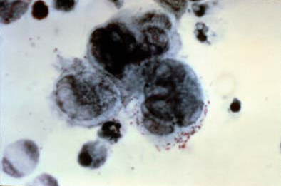
Muestra células gigantes multinucleadas.
Frotis de Tzanck con 3 células gigantes multinucleadas
Imagen: “Positive Tzanck test, showing three multinucleated giant cells in center” por NIAID. Licencia: Dominio Público
↓ Desenvolvimiento de virusVirusViruses are infectious, obligate intracellular parasites composed of a nucleic acid core surrounded by a protein capsid. Viruses can be either naked (non-enveloped) or enveloped. The classification of viruses is complex and based on many factors, including type and structure of the nucleoid and capsid, the presence of an envelope, the replication cycle, and the host range. Virology
Prevenir las complicaciones
Opciones:
Aciclovir
Valaciclovir
FamciclovirFamciclovirAn aminopurine derivative and prodrug of penciclovir which is a competitive inhibitor of herpes simplex 2 DNA polymerase. It is used to treat herpes simplex virus infection.Antivirals for Herpes Virus
El tratamiento debe ser administrado:
≤ 72 horas después del inicio de losLOSNeisseria síntomas
Durante la erupción de nuevas lesiones
EnENErythema nodosum is an immune-mediated panniculitis (inflammation of the subcutaneous fat) caused by a type IV (delayed-type) hypersensitivity reaction. It commonly manifests in young women as tender, erythematous nodules on the shins.Erythema Nodosum todos losLOSNeisseria pacientes inmunodeprimidos
Medidas de soporte
Analgésicos:
Acetaminofén
AINE
Antidepresivos tricíclicos
Gabapentinoides:
Gabapentina
Pregabalina
Corticosteroides (por ejemplo, prednisona) para el dolorDolorInflammation intenso o las complicaciones neurológicas
Antibióticos:
EnENErythema nodosum is an immune-mediated panniculitis (inflammation of the subcutaneous fat) caused by a type IV (delayed-type) hypersensitivity reaction. It commonly manifests in young women as tender, erythematous nodules on the shins.Erythema Nodosum caso de sospecha de infección bacteriana secundaria
Proporcionan cobertura estafilocócica y estreptocócica.
Tratamiento de enfermedades complicadas
Neuralgia postherpética:
LosLOSNeisseria antidepresivos tricíclicos y losLOSNeisseria gabapentinoides son la terapia de primera línea
Medicamentos tópicos (capsaicina) para controlar el dolorDolorInflammation leve o moderado
Opioides o glucocorticoides intratecales solo si fallan otras medidas
Herpes zóster oftálmico:
Pacientes inmunocompetentes: antivirales orales
Pacientes inmunodeprimidos: aciclovir IV
Considerar fuertemente la derivación a oftalmología temprana
NecrosisNecrosisThe death of cells in an organ or tissue due to disease, injury or failure of the blood supply.Ischemic Cell Damage retiniana aguda:
Indicada enENErythema nodosum is an immune-mediated panniculitis (inflammation of the subcutaneous fat) caused by a type IV (delayed-type) hypersensitivity reaction. It commonly manifests in young women as tender, erythematous nodules on the shins.Erythema Nodosum adultos inmunocompetentes ≥ 50 años de edad.
La vacuna recombinante contra el zóster también se recomienda enENErythema nodosum is an immune-mediated panniculitis (inflammation of the subcutaneous fat) caused by a type IV (delayed-type) hypersensitivity reaction. It commonly manifests in young women as tender, erythematous nodules on the shins.Erythema Nodosum adultos inmunodeprimidos ≥ 19 años de edad
Tipos de vacunas:
Vacuna viva contra el zóster (vacuna viva atenuada):
Ya no está disponible enENErythema nodosum is an immune-mediated panniculitis (inflammation of the subcutaneous fat) caused by a type IV (delayed-type) hypersensitivity reaction. It commonly manifests in young women as tender, erythematous nodules on the shins.Erythema Nodosum Estados Unidos, pero se utiliza enENErythema nodosum is an immune-mediated panniculitis (inflammation of the subcutaneous fat) caused by a type IV (delayed-type) hypersensitivity reaction. It commonly manifests in young women as tender, erythematous nodules on the shins.Erythema Nodosum otros países.
Contraindicada enENErythema nodosum is an immune-mediated panniculitis (inflammation of the subcutaneous fat) caused by a type IV (delayed-type) hypersensitivity reaction. It commonly manifests in young women as tender, erythematous nodules on the shins.Erythema Nodosum pacientes inmunodeprimidos
Vacuna recombinante contra el zóster (vacuna recombinante con glucoproteína E, p. ej., Shingrix)
Diagnóstico Diferencial
Herpes simple: Causado por el virusVirusViruses are infectious, obligate intracellular parasites composed of a nucleic acid core surrounded by a protein capsid. Viruses can be either naked (non-enveloped) or enveloped. The classification of viruses is complex and based on many factors, including type and structure of the nucleoid and capsid, the presence of an envelope, the replication cycle, and the host range. Virology del herpes simple y que se propaga por contacto directo con lesiones herpéticas o contacto con mucosas. La infección primaria suele presentarse con síntomas prodrómicos sistémicos seguidos de disuria, linfadenopatía dolorosa y grupos de vesículas dolorosas llenas de líquido sobre una base eritematosa. El diagnóstico se confirma con pruebas de laboratorio, como la PCRPCRPolymerase chain reaction (PCR) is a technique that amplifies DNA fragments exponentially for analysis. The process is highly specific, allowing for the targeting of specific genomic sequences, even with minuscule sample amounts. The PCR cycles multiple times through 3 phases: denaturation of the template DNA, annealing of a specific primer to the individual DNA strands, and synthesis/elongation of new DNA molecules.Polymerase Chain Reaction (PCR) y la AFD. El tratamiento incluye una terapia antiviralAntiviralAntivirals for Hepatitis B.
Celulitis: Infección bacteriana común de la piel que afecta a las capas más profundas de la dermisDermisA layer of vascularized connective tissue underneath the epidermis. The surface of the dermis contains innervated papillae. Embedded in or beneath the dermis are sweat glands; hair follicles; and sebaceous glands.Skin: Structure and Functions y el tejido subcutáneo. La causa más común de esta enfermedad es el Staphylococcus aureusStaphylococcus aureusPotentially pathogenic bacteria found in nasal membranes, skin, hair follicles, and perineum of warm-blooded animals. They may cause a wide range of infections and intoxications.Brain Abscess y el StreptococcusStreptococcusStreptococcus is one of the two medically important genera of gram-positive cocci, the other being Staphylococcus. Streptococci are identified as different species on blood agar on the basis of their hemolytic pattern and sensitivity to optochin and bacitracin. There are many pathogenic species of streptococci, including S. pyogenes, S. agalactiae, S. pneumoniae, and the viridans streptococci.Streptococcus pyogenes. La celulitis se presenta como una zona eritematosa y edematosa, caliente y sensible alALAmyloidosis tacto. El diagnóstico es clínico y el tratamiento consiste enENErythema nodosum is an immune-mediated panniculitis (inflammation of the subcutaneous fat) caused by a type IV (delayed-type) hypersensitivity reaction. It commonly manifests in young women as tender, erythematous nodules on the shins.Erythema Nodosum la administración de antibióticos y adaptarlos alALAmyloidosis agente etiológico sospechoso.
Foliculitis: Inflamación de losLOSNeisseria folículos pilosos causada por una infección bacteriana o fúngica. LosLOSNeisseria pacientes pueden presentar pápulas/pústulas eritematosas con prurito y dolorDolorInflammation alrededor de losLOSNeisseria folículos pilosos. El diagnóstico es clínico. El tratamiento suele ser de apoyo, pero enENErythema nodosum is an immune-mediated panniculitis (inflammation of the subcutaneous fat) caused by a type IV (delayed-type) hypersensitivity reaction. It commonly manifests in young women as tender, erythematous nodules on the shins.Erythema NodosumlosLOSNeisseria casos graves puede ser necesario un tratamiento antibiótico tópico u oral.
DermatitisDermatitisAny inflammation of the skin.Atopic Dermatitis (Eczema) alérgica de contacto: Inflamación de la piel debida alALAmyloidosis contacto con un alérgeno. LosLOSNeisseria pacientes suelen presentar una erupción local, roja, pruriginosa y pueden aparecer ampollas. El diagnóstico se realiza a través de losLOSNeisseria antecedentes clínicos y la exploración, pero puede utilizarse la prueba del parche cutáneo para determinar un factor desencadenante. El tratamiento incluye corticosteroides tópicos y evitar losLOSNeisseria alérgenos.
Molusco contagioso: Es una infección viral de la piel causada por un poxvirus que suele aparecer enENErythema nodosum is an immune-mediated panniculitis (inflammation of the subcutaneous fat) caused by a type IV (delayed-type) hypersensitivity reaction. It commonly manifests in young women as tender, erythematous nodules on the shins.Erythema Nodosum niños de < 5 años de edad. Las lesiones son pápulas agrupadas, de color carne, enENErythema nodosum is an immune-mediated panniculitis (inflammation of the subcutaneous fat) caused by a type IV (delayed-type) hypersensitivity reaction. It commonly manifests in young women as tender, erythematous nodules on the shins.Erythema Nodosum forma de cúpula, con umbilicación central. La enfermedad es leve enENErythema nodosum is an immune-mediated panniculitis (inflammation of the subcutaneous fat) caused by a type IV (delayed-type) hypersensitivity reaction. It commonly manifests in young women as tender, erythematous nodules on the shins.Erythema Nodosum pacientes inmunocompetentes y se resuelve por sí sola enENErythema nodosum is an immune-mediated panniculitis (inflammation of the subcutaneous fat) caused by a type IV (delayed-type) hypersensitivity reaction. It commonly manifests in young women as tender, erythematous nodules on the shins.Erythema Nodosum meses. Un paciente inmunodeprimido puede presentar lesiones extensas y enfermedad sistémica que requiere tratamiento. El diagnóstico es clínico. La crioterapia con nitrógeno líquido es la primera línea enENErythema nodosum is an immune-mediated panniculitis (inflammation of the subcutaneous fat) caused by a type IV (delayed-type) hypersensitivity reaction. It commonly manifests in young women as tender, erythematous nodules on the shins.Erythema NodosumlosLOSNeisseria pacientes que requieren terapia.
Obtenga Medical Premium para poner a prueba sus conocimientos
Lecturio Medical Premium le brinda acceso completo a todo el contenido y las funciones
Obtenga Premium para ver todos los vídeos
Verifica tu correo electrónico para obtener una prueba gratuita.
Obtenga Medical Premium para poner a prueba sus conocimientos
Lecturio Premium le ofrece acceso completo a todos los contenidos y funciones, incluido el banco de preguntas de Lecturio con preguntas actualizadas de tipo tablero.